SlideShare a Scribd company logo
Contrast Media
in Radiology
what is used and
why
Introduced by
Abdulrahman Alquait
Medical imaging MSc student
-What is a contrast medium or
contrast agent?
It is a substance used to
enhance the contrast of
structures or fluids within the
body in medical imaging¹.
¹ contrast agent at Dorland's Medical Dictionary
How can we achieve this?
 What type of imaging modality being
used?
 What part of the body being investigated?
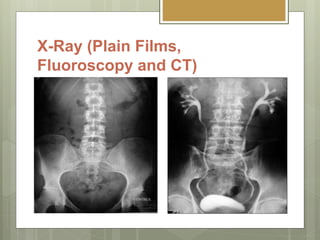
X-Ray (Plain Films,
Fluoroscopy and CT)
 Beam attenuation.
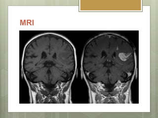
MRI
 T1 & T2 Relaxation times.
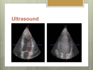
Ultrasound
 Decreasing reflection or increasing
backscatter of echoes.
Types of contrast media in
Radiography.
 Positive Contrast Agents.
 High atomic number.
 (e.g. iodine(53) and Barium(56))
 Negative Contrast Agents.
 Low atomic number.
 (e.g. carbon dioxide (CO2) and air )
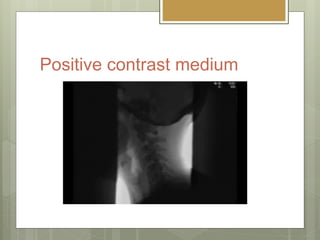
Positive contrast medium
1. Water insoluble.
 Barium sulphate.
 Barium meals and barium enemas to study
the GI tract.
Positive contrast medium
2. Water soluble.
 Iodine compounds.
 Intravascular injections.
Positive contrast medium
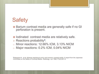
Safety
 Barium contrast media are generally safe if no GI
perforation is present.
 Iodinated contrast media are relatively safe.
 Reactions probability²:
o Minor reactions: 12.66% ICM, 3.13% NICM
o Major reactions: 0.2% ICM, 0.04% NICM
²Katayama H, et al. Adverse reactions to ionic and nonionic contrast media. A report from the Japanese
Committee on the Safety of Contrast Media. Radiology. Jun 1990;175(3):621-8
More Related Content

Contrast Media in Radiology

  • 1. Contrast Media in Radiology what is used and why Introduced by Abdulrahman Alquait Medical imaging MSc student
  • 2. -What is a contrast medium or contrast agent? It is a substance used to enhance the contrast of structures or fluids within the body in medical imaging¹. ¹ contrast agent at Dorland's Medical Dictionary
  • 3. How can we achieve this?  What type of imaging modality being used?  What part of the body being investigated?
  • 4. X-Ray (Plain Films, Fluoroscopy and CT)  Beam attenuation.
  • 5. MRI  T1 & T2 Relaxation times.
  • 6. Ultrasound  Decreasing reflection or increasing backscatter of echoes.
  • 7. Types of contrast media in Radiography.  Positive Contrast Agents.  High atomic number.  (e.g. iodine(53) and Barium(56))  Negative Contrast Agents.  Low atomic number.  (e.g. carbon dioxide (CO2) and air )
  • 8. Positive contrast medium 1. Water insoluble.  Barium sulphate.  Barium meals and barium enemas to study the GI tract.
  • 10. Positive contrast medium 2. Water soluble.  Iodine compounds.  Intravascular injections.
  • 12. Safety  Barium contrast media are generally safe if no GI perforation is present.  Iodinated contrast media are relatively safe.  Reactions probability²: o Minor reactions: 12.66% ICM, 3.13% NICM o Major reactions: 0.2% ICM, 0.04% NICM ²Katayama H, et al. Adverse reactions to ionic and nonionic contrast media. A report from the Japanese Committee on the Safety of Contrast Media. Radiology. Jun 1990;175(3):621-8