Bacterial morphology
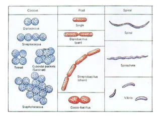
- 2. Introduction • Bacteria belong to the kingdom Monera. They are unicellular organisms • They are classified according to three shapes 1. Spherical (cocci) 2. Rod (bacillus) 3. Spiral (spirillum)
- 5. Spherical (cocci) E.g. Staphoolococcus aureus Causes pneumonia
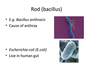
- 6. Rod (bacillus) • E.g. Bacillus anthracis • Cause of anthrax • Escherichia coli (E.coli) • Live in human gut
- 7. Spirillum (spiral) • E.g.Treponema pallidum • Causes syphilis
- 8. Bacterial cell structure • Surface layers - capsule, cell wall, cell membrane • Cytoplasm - nuclear material, ribosome, mesosome, inclusion bodies etc. • Appendages - fllagella, pili or fimbriae • Special structure - endospore
- 10. Bacterial cell
- 11. Surface layers 1. Capsule. 2. Cell wall. 3. Cell membrane.
- 12. 1. Capsule and Slime layer These are structures surrounding the outside of the cell envelope. They usually consist of polysaccharide; however, in certain bacilli they are composed of a polypeptide (polyglutamic acid). They are not essential to cell viability and some strains within a species will produce a capsule, whilst others do not. Capsules are often lost during in vitro culture. Functions: Attachment Protection from phagocytic engulfment. Resistance to drying. Depot for waste products. Reservoir for certain nutrients. Protection.
- 13. Capsule
- 14. 2. Cell wall Next lecture
- 15. 3. Cell Membrane Function: a. Control permeability b. Transport electrons and protons for cellular metabolism. c. Contain enzymes to synthesis and transport solute into the cell. d. Secret hydrolytic enzymes e. Site of biosynthesis of DNA, cell wall polymers and membrane lipids. f. Electron transport and oxidative phosphorylation.
- 16. Cell membrane
- 17. Functions of cell membrane
- 18. Functions of cell membrane
- 19. Cytoplasm • Composed largely of water (80%). 1. Nuclear material. 2. Ribosome. 3. Mesosome. 4. Plasmids 5. Inclusion bodies etc.
- 20. 1.Nucleic material • Lacking nuclear membrane, absence of nucleoli, hence known as nucleic material or nucleoid, one to several per bacterium. • A single large circular double stranded DNA no histone proteins. The only proteins associated with the bacterial chromosomes are those for DNA replication, transcription etc.
- 22. 2. Ribosome • Numerous in number. • 15-20nm in diameter withwith70S. • Distributed throughout the cytoplasm. • Sensitive to streptomycin and erythromycin site of protein synthesis.
- 24. 3. Mesosome • Mesosomes are specialized structures formed by convoluted invaginations of cytoplasmic membrane. • Divided into septal and lateral mesosome. • Irregular in shape. Functions a. Increase in membrane surface, which may be useful as a site for enzyme activity in respiration and transport. b. May participate in cell replication by serving as a place of attachment for the bacterial chromosome
- 26. 4. Plasmids • Plasmids are small, circular, extrachromosomal , double-stranded DNA molecule. • They are capable of self-replication. • They contain genes that confer some properties, such as antibiotic resistance, virulence factor. • Plasmids are not essential for cellular survival.
- 27. 5.Inclusion bodies Many bacteria have a variety of granules in their cytoplasm. These granules are store deposits of nutrients like phosphate, sulphur and fats etc. Examples Granules of various kinds: * Glycogen granules, they can be stained with iodine, as they are polysaccharides. *poly B- hydroxybutyrate inclusion bodies (PHB) are carbon reservoirs, providing energy for biosynthesis. * Volutin granules (metachromatic granules) are made up of polyphosphate. in general, starvation of cell for almost any nutrients leads to the formation of this to serve as an intracellular phosphate reservoir.
- 28. Appendages The filamentous structures protruding from the cell wall of a bacterial cell are called appendages. These are of two types 1. Flagella 2. Pili or fimbriae
- 29. 1. Flagella • Some bacterial species are mobile and possess locomotory organelles - flagella. • The diameter of a flagellum is thin, 20 nm, and long with some having a length 10 times the diameter of cell. Due to their small diameter, flagella cannot be seen in the light microscope. • Bacteria can have one or more flagella arranged in clumps or spread all over the cell.
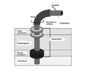
- 30. Ultra structure of Flagella • Flagella consists of repeating units of a protein------- flagellin. • Flagellin molecules assemble to form fibers. • A flagellum is formed when three fibers twist to form a helix. • Flagellum is made up of three parts (Electron Micrography). 1. Filament 2. Hook 3. Basal body
- 31. Filament
- 32. The structure of the bacterial flagella allows it to spin like a propeller and thereby propel the bacterial cell; clockwise or counter clockwise. – Bacterial flagella provides the bacterium with mechanism for swimming toward or away from chemical stimuli, a behavior known as Chemotaxix, chemosenors in the cell envelope can detect certain chemicals and signal the flagella to respond.
- 33. The number and arrangements of flagella are variable. Monotrichous. One flagella from one end. (Vibrio cholerae) Lophotrichous. Tuft of flagella at one pole. (Pseudomonas) Amphitrichous. Flagella at both ends. (spirillum serpens) Peritrichous. Flagella arise from all over the surface. (E. coli)
- 34. 2. Pili or Fimbriae • Shorter, straighter, thinner, non helical and numerous than flagella. • Mostly on gram - bacteria. • They are usually found in large numbers over the cell surface, arising from cell membrane. • Structure: They have a hollow core and are composed of proteins which can be dissociated into smaller units Pilin . It belongs to a class of protein Lectin which bond to cell surface polysaccharide.
- 35. Function a. Conjugation: Specialized sex pilus or F pilus serves for the attachment of two bacterial cell before conjugation and act as conjugation tube. b. Adhesion: Serves as attachment structures or adhesins. E.g. E.coli have pili that help them to adhere to intestinal lining and causes diarrhea. c. Receptor site: Act as site for some bacteriophages. Bacteriophages attaches to pili and transfer their genetic information to bacterial cell.
- 36. Special Structure-----Endospore Dormant cell. Produced when starved. Resistant to adverse conditions, ultraviolet radiation, high temperatures, extreme freezing and chemical disinfectants. Organic solvents. Contain calcium dipicolinate DPA, dipicolinic acid. Mostly gram positive bacteria (Bacillus and clostridium).
- 37. Structure and chemical composition • Core • Dipicolinic acid • Spore Cortex • Spore coat • Exosporium
- 38. • Core: It is the centre of the spore that contains the chromosomes, all components of proteins synthesizing machinery, enzymes etc. • Dipicolinic acid (DPA): It is a spore-specific chemical that appears to help in the ability for endospores to maintain dormancy. This chemical comprises up to 10-15% of the spore's dry weight. It is present in association with high amounts of calcium in the core. The heat resistance of the endospore is due to Calcium dipicolinate. • Spore wall: Inner most lining surrounding the core. It consists of Peptidoglycan and becomes cell wall of germinating vegetative cells. • Cortex: Thickest layer of core envelope.
- 39. • Spore coat: The outer most impermeable layer which confers resistance to the spore to antibacterial chemical agents. • Exosporium: It is the outer most membrane that contains lipoproteins and some carbohydrates.
- 40. Detailed steps in endospore formation


