SlideShare a Scribd company logo
Dr SHILPA JOY
V S Dental College
13/1/2017 99
CONTENTS
 Introduction
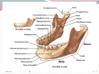
 Anatomy of mandible
 Evolution of mandible
 Mandibular growth mechanisms-Brief history
 Prenatal growth of mandible
 Postnatal development
 Growth progression-mechanism & site
 Age changes in mandible
 Theories of mandibular growth
 Problems of mandibular growth and orthodontic
significance
 Conclusion
 References 23/1/2017 99
INTRODUCTION
The human mandible has no one design for life.Rather it
adapts and remodels through the seven stages of life,from the
slim arbiter of things to come in the infant,through a powerful
dentate machine and even weapon in the full flesh of
maturity ,to the pencil thin,porcelain like problem that we
struggle to repair in the adversity of old age –D.E POSWILLO
It is attached only by ligaments and muscles to immovable
bones of the skull.
The temporomandibular or ginglymo-diarthroidal joints are
the only visible movable articulations in the head. The rest of
the bones of the skull move in union when the head is moved
as a whole.
33/1/2017 99
DEFINITIONS RELATED TO GROWTH
 Moss: change in morphological parameters which is measurable.
 Moyers: Quantitative aspect of biological development per unit of
time.
 Todd: An increase in size.
 Krogman: increase in size change in proportions and progressive
complexity
 J.S. Huxley: The self multiplications of living substance
 Meridith:entire series of sequential anatomic and Physiologic
changes taking place from beginning of pre natal life to serenity
DEVELOPMENT
 Todd:progress towards maturity
 Moyers:All naturally occuring unidirectional changes in the life of
an individual from its existence as a single cell to its elaboration as a
multifunctional unit terminating in death
43/1/2017 99
ANATOMY OF MANDIBLE
53/1/2017 99
63/1/2017 99
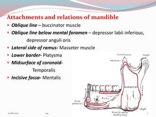
Attachments and relations of mandible
 Oblique line – buccinator muscle
 Oblique line below mental foramen – depressor labii inferious,
depressor anguli oris
 Lateral side of ramus- Masseter muscle
 Lower border- Platysma
 Midsurface of coronoid-
Temporalis
 Incisive fossa- Mentalis
73/1/2017 99
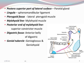
 Postero superior part of lateral surface – Parotid gland
 Lingula – sphenomandibular ligament
 Pterygoid fossa - lateral pterygoid muscle
 Mylohyoid line- Mylohyoid muscle
 Posterior end of mylohyoid line-
superior constrictor muscle
 Digastric fossa- Anterior belly
of digastric
 Genial tubercle -Genioglossus &
Geniohyoid
83/1/2017 99
EVOLUTION OF MANDIBLE
 The agnatha, the earliest type of
vertebrate, had its mouth opening on
the ventral side anteriorly along the
vertebral axis. They did not have jaws
 The Placoderms, had 7 arches.The first
arch was lost.Their new first arch
became the Mandibular arch that
formed the jaws.The upper half of
mand.arch became palatoquadrate cartilage
and lower half became the mandibular
or meckels cartilage
 In Elasmobrancs,the jaws were formed by
mandibular arch (arch 1) & hyoid arch(arch 2)
The upper half of hyoid arch became
hyomandibular ligament and lower half
became hyoid cartilage 93/1/2017 99
In amphibians the hyomandibular
ligament became the stapes .They had
a dentary bone in the anterior end of
the original cartilaginous jaw. At its
posterior extremity it articulated with
the quadrate bone
In reptiles like early synapsid ,that gave rise to mammals ,the jaw joint is
formed by the articular(lower) & quadrate(upper) bones.The joint was a
simple hinge at the posterior of jaw
In mid and late synapsid reptiles,the dentary bone (lower jaw) increase in
size as muscle and bite force increased,but force on the joint decreased as
the muscle insertion point shifted to allow greater jaw mobility
The articular and quadrate bones at the jaw joint became smaller and was
loosely attached with the dentary
103/1/2017 99
 The coronoid process of dentary bone formed to accommodate these
changing forces
 Ultimately in mammals,the jaw joint shifted from a articular-quadrate
joint to a dentary-squamosal joint.the condylar process formed to create
a new articulating surface
113/1/2017 99
MANDIBULAR GROWTH PATTERNS- A HISTORY
• HUNTER (1771) compared a series of dried mandibles and concluded that in
order to attain space for permanent molar teeth the mandible must grow by
posterior apposition of ramus accompanied by anterior ramus resorption.
• HUMPHRY (1866) studied growth of mandible by inserting metal rings in
the anterior and posterior margins of mandibular ramus in growing pig.Rings
placed on posterior border became more deeply embedded but rings placed
on anterior surface were released
• BRASH (1924) fed pigs the madder plant root(alizarin ) which labeled
appositional growth
 WEINMAN AND SICHER (1940) with the help of longitudinal cephalometrics
and evidence from experiments of animals, focused attention on the
mandibular condyle as a major factor in growth of the mandible.
A STUDY OF POSTNATAL GROWTH OF HUMAN MANDIBLE-DONALD H ENLOW,DAVID B HARRIS
AJODO,JAN 1964
123/1/2017 99
 BRODIE believed that superior and posterior growth of the
condyle along with apposition of the posterior border of the
ramus and alveolar border resulted in development of
mandible.
 RICKETTS(1950)by superimpositions on lower border of the
mandible showed that the condyle followed a superior and
posterior course. He also noted that the mandibular growth
was not same and that the relationship of the mandibular
plane to the Frankfort Horizontal plane was changing about
one degrees every 3 yrs in a typical facial pattern.
 MOSS(1960) envisioned the growth of the mandible as a
logarithmic spiral constructed via the path of the mandibular
nerve.
133/1/2017 99
 BJORK (1963 )conducted a study with tantalum implants and
suggested that
1. Growth in length of the mandible occurs at the condyles.
2. The anterior aspect of chin is extremely stable
3. The thickening of the symphysis takes place by appostion on its
posterior surface and lower border which contributes to
increase in height of symphysis.
4. At the region of the condyles there is upward and forward
curving growth.
5. The mandibular canal is not remodeled and the trabaculae
related to the canal are stationary. Hence the curvature of the
mandibular canal generally reflects the earlier shape of the
mandible
VARIATION IN GROWTH PATTERN OF HUMAN MANDIBLE;A LONGITUDINAL RADIOGRAPHIC
STUDY BY IMPLANT METHOD-ARNE BJORK ;J DENT RES 1963
.
143/1/2017 99
Growth & development of an individual can be divided in to
Pre-natal
Post-natal
Period of Ovum
1-14th day
Period of embryo
14th – 56th day
Period of Fetus
56th – 270th day
153/1/2017 99
PRENATAL GROWTH
PERIOD OF OVUM
Fertilization-Ampulla of uterine tube
zygote
mitosis
Cluster of cells(Blastomere)
mitosis
Morula(16 cell structure)
Blastocyst
163/1/2017 99
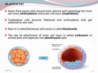
BLASTOCYST
Some fluid passes into morula from uterine wall seperating the inner
cell mass (embryoblast) and outer cell mass (trophoblast)
Trophoblast cells become flattened and embryoblast cells get
attached to one side
Now it is called blastocyst and cavity is called blastocoele
The site of attachment of inner cell mass is called embryonic or
animal pole and opposite site abembryonic pole
MORULA
173/1/2017 99
PERIOD OF EMBRYO
PRESOMITE PERIOD(8-20th day)
 Trophoblastic layer differentiate into synctiotrophoblast and
cytotrophoblast layers
SYNCTIOTROPHOBLAST- Outer cells that invades
endometrium and its vessels to establish maternal blood
circulation to developing embryo-UTEROPLACENTAL
CIRCULATION
PRESOMITE SOMITE POSTSOMITE
183/1/2017 99
INNER CELL MASS
Differentiation
BILAMINAR DISC
BLASTOCYSTIC CAVITY is now called as primitive yolk sac
AMNIOTIC CAVITY develops between epiblast and cytotrophoblast
HYPOBLAST
Squamous
/cuboidal
EPIBLAST
Columnar
cells
193/1/2017 99
 Extra embryonic mesoderm(EEM)
formed of loose connective tissue ,
differentiate between developing
embryo and cytotrophoblast
 Chorionic cavity is formed by
fusion of number of lacunae that
develop in EEM
 Expansion of chorionic cavity reduces size of primitive yolk
sac,forming secondary yolk sac, occurs by end of second week
 3rd week- Gastrulation occurs
Bilaminar disc-Trilaminar disc
203/1/2017 99
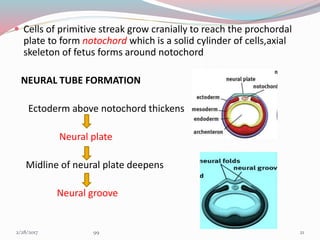
 Cells of primitive streak grow cranially to reach the prochordal
plate to form notochord which is a solid cylinder of cells,axial
skeleton of fetus forms around notochord
NEURAL TUBE FORMATION
Ectoderm above notochord thickens
Neural plate
Midline of neural plate deepens
Neural groove
213/1/2017 99
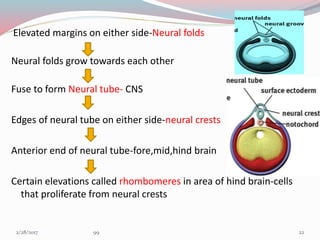
Elevated margins on either side-Neural folds
Neural folds grow towards each other
Fuse to form Neural tube- CNS
Edges of neural tube on either side-neural crests
Anterior end of neural tube-fore,mid,hind brain
Certain elevations called rhombomeres in area of hind brain-cells
that proliferate from neural crests
223/1/2017 99
NEURAL CREST CELLS
Forms from neuro ectoderm
Migrate & differentiate extensively with in the developing embryo
Spinal & cranial sensory ganglia, Sympathetic neurons, Schwann cells, pigment
cells & meninges
Most of the connective tissue of the head is formed
Migration is essential for development of teeth & face
All the tissues of teeth (except enamel) & its supporting apparatus are derived
directly from these cells
233/1/2017 99
SOMITE PERIOD(21ST -31ST day of IUL)
 Rapid growth of cranial end of embryo,caudal end lags
behind- CEPHALOCAUDAL GRADIENT OF GROWTH
Head-1/2 of total embryonic disk length
BRANCHIAL /PHARYNGEAL ARCHES
 In specific areas,the migrating and rapidly proliferating
ectomesenchyme cells develops elevation between ectoderm and
endoderm
 4th week of IUL Elevations seen in ventral foregut
5th arch perishes Formation of 6 pharyngeal arches
(bilaterally)
Finally 5 arches remain
243/1/2017 99
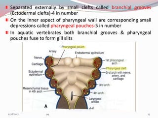
Separated externally by small clefts called branchial grooves
(Ectodermal clefts)-4 in number
On the inner aspect of pharyngeal wall are corresponding small
depressions called pharyngeal pouches-5 in number
In aquatic vertebrates both branchial grooves & pharyngeal
pouches fuse to form gill slits
253/1/2017 99
263/1/2017 99
DEVELOPMENT OF MANDIBLE
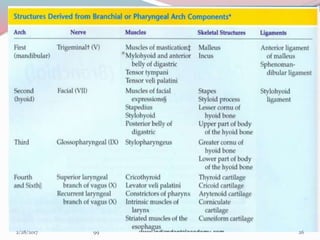
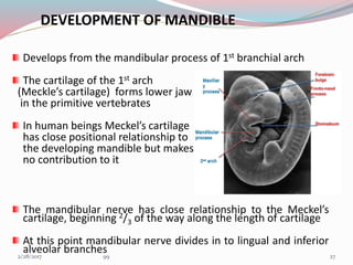
Develops from the mandibular process of 1st branchial arch
The cartilage of the 1st arch
(Meckle’s cartilage) forms lower jaw
in the primitive vertebrates
In human beings Meckel’s cartilage
has close positional relationship to
the developing mandible but makes
no contribution to it
The mandibular nerve has close relationship to the Meckel’s
cartilage, beginning 2/3 of the way along the length of cartilage
At this point mandibular nerve divides in to lingual and inferior
alveolar branches 273/1/2017 99
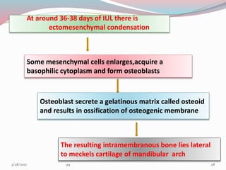
At around 36-38 days of IUL there is
ectomesenchymal condensation
Some mesenchymal cells enlarges,acquire a
basophilic cytoplasm and form osteoblasts
Osteoblast secrete a gelatinous matrix called osteoid
and results in ossification of osteogenic membrane
The resulting intramembranous bone lies lateral
to meckels cartilage of mandibular arch
283/1/2017 99
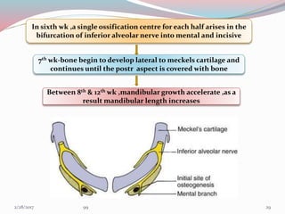
In sixth wk ,a single ossification centre for each half arises in the
bifurcation of inferior alveolar nerve into mental and incisive
7th wk-bone begin to develop lateral to meckels cartilage and
continues until the postr aspect is covered with bone
Between 8th & 12th wk ,mandibular growth accelerate ,as a
result mandibular length increases
293/1/2017 99
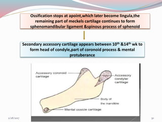
Ossification stops at apoint,which later become lingula,the
remaining part of meckels cartilage continues to form
sphenomandibular ligament &spinous process of sphenoid
Secondary accessory cartilage appears between 10th &14th wk to
form head of condyle,part of coronoid process & mental
protuberance
303/1/2017 99
FATE OF MECKELS CARTILAGE
Posterior extremity forms malleus, incus &
sphenomandibular ligament
Most of the cartilage is absorbed except for some
portion in midline which may cause endochondral
ossification
FETAL PERIOD
Endochondral bone formation seen only in 3 areas
• Condylar process
• Coronoid process
• Mental region
313/1/2017 99
Condylar process
 About 5th week of I.U.L. area of
mesenchymal condensation above
the ventral part of developing mandible
 About 10th wk develops into cone shaped
cartilage
 By 14th week starts ossification
 By 4 months migrates inferiorly and fuses with ramus
 4th month onwards replaced by bone but proximal end persists into
adulthood acting as
Growth cartilage & Articular cartilage
 Condylar head separated from temporal bone by thin disc of connective
tissue – future articular disc
323/1/2017 99
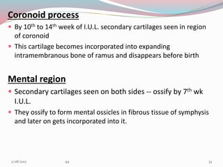
Coronoid process
 By 10th to 14th week of I.U.L. secondary cartilages seen in region
of coronoid
 This cartilage becomes incorporated into expanding
intramembranous bone of ramus and disappears before birth
Mental region
 Secondary cartilages seen on both sides -- ossify by 7th wk
I.U.L.
 They ossify to form mental ossicles in fibrous tissue of symphysis
and later on gets incorporated into it.
333/1/2017 99
POST NATAL DEVELOPMENT
MANDIBLE AT BIRTH
MANDIBULAR
GROWTH-FIRST YEAR
GROWTH
PROGRESSION AFTER
FIRST YEAR-
MECHANISM & SITE
343/1/2017 99
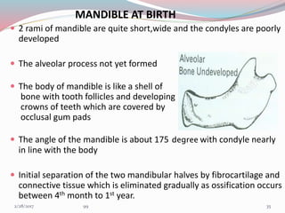
MANDIBLE AT BIRTH
 2 rami of mandible are quite short,wide and the condyles are poorly
developed
 The alveolar process not yet formed
 The body of mandible is like a shell of
bone with tooth follicles and developing
crowns of teeth which are covered by
occlusal gum pads
 The angle of the mandible is about 175 degree with condyle nearly
in line with the body
 Initial separation of the two mandibular halves by fibrocartilage and
connective tissue which is eliminated gradually as ossification occurs
between 4th month to 1st year.
353/1/2017 99
MANDIBULAR GROWTH DURING FIRST YEAR
Appositional growth especially active at
 Alveolar border
 Distal and superior surface of ramus
 Condyle
 Lower border of mandible
 Lateral surface of mandible
 After first year growth becomes more selective,condyle shows
considerable activities , mandible moves and grows forward and
downward
363/1/2017 99
GROWTH PROGRESSION-MECHANISM & SITE
MANDIBULAR CONDYLE
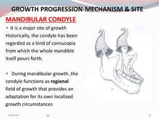
 It is a major site of growth
Historically, the condyle has been
regarded as a kind of cornucopia
from which the whole mandible
itself pours forth.
 During mandibular growth ,the
condyle functions as regional
field of growth that provides an
adaptation for its own localized
growth circumstances
373/1/2017 99
 The condylar cartilage is a secondary type of cartilage
 Its real contribution is to provide regional adaptive growth
 Main functional role of condyle is
(1) provides a pressure tolerant articular contact
(2) it makes possible a multidimensional growth capacity in
response to ever-changing, developmental conditions and
variations.
 The condylar growth mechanism itself is a clear-cut process.
Cartilage is a special non-vascular tissue and is involved
because variable levels of compression occur at its articular
contact with temporal bone
383/1/2017 99
 An endochondral growth mechanism is required ,because the
condyle grow in a direction towards its articulation in the face of
direct pressure
 Intramembranous type couldn’t operate because the periosteal
mode of osteogenesis is not pressure adapted
 In Figure the endochondral bone tissue (b) , formed in association
with the condylar cartilage (a) is laid down only in medullary portion
 The enclosing bony cortices (c) are produced by periosteal-
endosteal osteogenic activity
393/1/2017 99
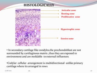
HISTOLOGICALLY
Articular zone
Resting zone
Proliferative zone
Hypertrophic zone
Erosive zone
• In secondary cartilage like condyles,the prechondoblast are not
surrounded by cartilaginous matrix ,thus they are exposed to
environment and are moldable to external influences
•Codylar cellular arrangement is multidirectional unlike primary
cartilage where its arranged in rows
403/1/2017 99
 Anterior margin of condylar neck is
depository, this surface is part of sigmoid notch
 Posterior edge which grades into posterior
border of ramus is also depository
The lingual and buccal sides of neck characteristically have a
resorptive surface. This is because condyle is quite broad and
neck is narrow
413/1/2017 99
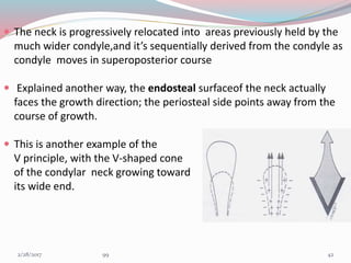
 The neck is progressively relocated into areas previously held by the
much wider condyle,and it’s sequentially derived from the condyle as
condyle moves in superoposterior course
 Explained another way, the endosteal surfaceof the neck actually
faces the growth direction; the periosteal side points away from the
course of growth.
 This is another example of the
V principle, with the V-shaped cone
of the condylar neck growing toward
its wide end.
423/1/2017 99
What is the physical force that produces the forward and
downward primary displacement of mandible ???????
For many years it was presumed that
 Cartilage is pressure adapted type of tissue & creates a thrust of
mandible against its articular bearing surface
 proliferation of cartilage towards its contact thereby pushes the whole
mandible away from it.
But,Bilaterally condyle- lacking mandibles occupy an essentially
normal anatomic position and the mandible functions in
movements
These observations suggested 2 conclusions.
1. Condyles may not play the kingpin role of a “master center”.
2. The whole mandible can become displaced anteriorly and
inferiorly into its functional position without a "push" against the
basicranium
433/1/2017 99
 The current thinking is that condylar cartilage does have a
measure of intrinsic genetic programming .
 The cartilage cells are coded and geared to divide ,but
extracondylar factors are needed to sustain this activity
 So overall mandibular length can be clinically increase or
decrease depending on class II or class III if this were done during
period of active condylar growth
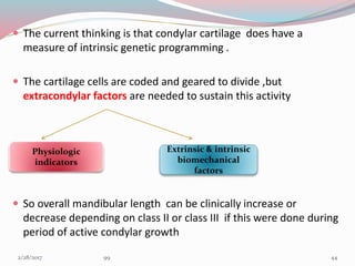
Physiologic
indicators
Extrinsic & intrinsic
biomechanical
factors
443/1/2017 99
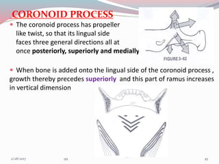
CORONOID PROCESS
 The coronoid process has propeller
like twist, so that its lingual side
faces three general directions all at
once posteriorly, superiorly and medially.
 When bone is added onto the lingual side of the coronoid process ,
growth thereby precedes superiorly and this part of ramus increases
in vertical dimension
453/1/2017 99
 These same deposits of bone on the
lingual side also bring about a
posterior direction of growth
movement .
 Produces backward movement of
two coronoid process even though
deposits on the inside (lingual) surface.
 These same deposits on the lingual
side also bring about medial direction
of growth in order to lengthen corpus
 Area occupied by anterior part of ramus
in mandible 1 becomes relocated and
remodeled into posterior part of corpus
in mandible 2.
463/1/2017 99
CORPUS / BODY OF MANDIBLE
 Outer surface-depository & medial surface(inferior aspect)-
Resorptive,remodelling is in the form of ‘L’
 Depository area -from the superior half of medial surface of
corpus to anterior half of medial surface of ramus(below coronoid)
 Resorptive area –from inferior half of medial surface of corpus to
posterior half of medial surface of ramus (below condyle)
473/1/2017 99
RAMUS
 At birth the two rami of mandible are
quite short, they grow by the process
of direct surface apposition and
remodeling.
 THE PRINCIPLE GROWTH VECTORS ARE IN POSTERIOR & SUPERIOR
DIRECTION
 Resorption occurs on the anterior
surface of ramus while bone deposition
occurs on posterior surface.
 Bone growth occurs at the mandibular condyle and along the
posterior part of ramus to the same extent as anterior part has
undergone resorption
483/1/2017 99
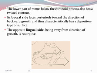
 The lower part of ramus below the coronoid process also has a
twisted contour.
 Its buccal side faces posteriorly toward the direction of
backward growth and thus characteristically has a depository
type of surface.
 The opposite lingual side, being away from direction of
growth, is resorptive.
493/1/2017 99
IMPORTANCE OF RAMUS
 It positions the lower arch in occlusion with the upper.
 It is continuously adaptive to the multitude of changing
craniofacial conditions.
 Attach the mastication muscle and must accommodate the
increasing mass of masticatory muscle inserted into it.
 Bridges the pharyngeal compartment.
 The horizontal breadth of ramus determines the
anteroposterior positioning of lower arch.
 Height of ramus accommodates the vertical dimension and
growth of nasal and masticatory components of face.
 Remodeling and relocation give space to accommodate
erupting permanent molar.
503/1/2017 99
RAMUS UPRIGHTING
 Greater amounts of bone additions
on the inferior part of the posterior
border than on the superior part.
 A correspondingly greater amount
of matching resorption on the anterior
border takes place inferiorly than
superiorly.
 A "remodeling" rotation of ramus
alignment thus occurs.
 In diagram the pharynx enlarges
horizontally from a to a’ .
 The ramus enlarges correspondingly from b to b’
 Angle c is reduced to c’ to accommodate the vertical
increase,which allows for considerable extent of vertical
nasomaxillary growth 513/1/2017 99
 Vertical lengthening of the ramus continues to take place after
horizontal ramus growth slows or ceases
 Resorption takes place on the upper part
of the posterior border.
 A forward growth direction can occur
on the anterior border in the upper part
of the coronoid process.
 A posterior direction of remodeling takes place in the lower part of the
posterior border,this result in more upright alignment and longer vertical
dimension of ramus without increase in breadth
In fig mandible a is superimposed
over b remodeling changes outlined
above serve simply to alter the ramus
angle without increasing its breadth
523/1/2017 99
• The growth and remodeling changes of both ramus and middle cranial
fossa produces lowering of mandibular arch .This accommodate vertical
expansion of nasomaxillary complex .
• A vertical imbalance thus occurs ,this ‘opens’ anterior bite,only the first
and second molars are in occlusal contact
• The amount of upward mandibular tooth drift is much less than the
downward drift and displacement of maxillary teeth.
• This is one of the several reason why
orthodontic purpose, often attack maxillary
dentition ,eventhough given malocclusion
can be largely based on improper positioning
of mandible
533/1/2017 99
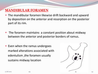
MANDIBULAR FORAMEN
 The mandibular foramen likewise drift backward and upward
by deposition on the anterior and resorption on the posterior
part of its rim.
 The foramen maintains a constant position about midway
between the anterior and posterior borders of ramus.
 Even when the ramus undergoes
marked alterations associated with
edentulism ,the foramen usually
sustains midway location
543/1/2017 99
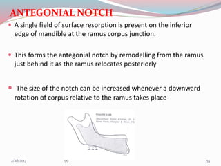
ANTEGONIAL NOTCH
 A single field of surface resorption is present on the inferior
edge of mandible at the ramus corpus junction.
 This forms the antegonial notch by remodelling from the ramus
just behind it as the ramus relocates posteriorly
 The size of the notch can be increased whenever a downward
rotation of corpus relative to the ramus takes place
553/1/2017 99
ANTEGONIAL NOTCH –CLINICAL
SIGNIFICANCE
 Deep notched subjects have retrusive mandible with
shorter corpus, less ramus height and increase gonial
angle.
 Mandibular growth directions in deep notched patients
were more vertically directed as measured by facial axis
and the mandibular plane angle.
 Deep notched subjects had longer total facial height and
longer lower facial height,Smaller saddle angle
 Deep notch patients required a longer duration of
orthodontic treatment.
563/1/2017 99
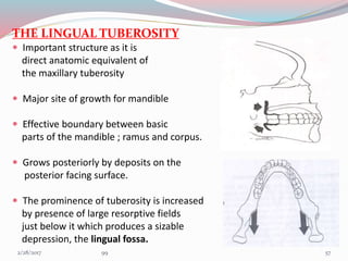
THE LINGUAL TUBEROSITY
 Important structure as it is
direct anatomic equivalent of
the maxillary tuberosity
 Major site of growth for mandible
 Effective boundary between basic
parts of the mandible ; ramus and corpus.
 Grows posteriorly by deposits on the
posterior facing surface.
 The prominence of tuberosity is increased
by presence of large resorptive fields
just below it which produces a sizable
depression, the lingual fossa.
573/1/2017 99
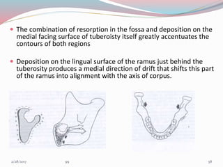
 The combination of resorption in the fossa and deposition on the
medial facing surface of tuberoisty itself greatly accentuates the
contours of both regions
 Deposition on the lingual surface of the ramus just behind the
tuberosity produces a medial direction of drift that shifts this part
of the ramus into alignment with the axis of corpus.
583/1/2017 99
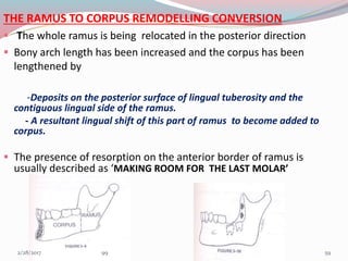
THE RAMUS TO CORPUS REMODELLING CONVERSION
 The whole ramus is being relocated in the posterior direction
 Bony arch length has been increased and the corpus has been
lengthened by
-Deposits on the posterior surface of lingual tuberosity and the
contiguous lingual side of the ramus.
- A resultant lingual shift of this part of ramus to become added to
corpus.
 The presence of resorption on the anterior border of ramus is
usually described as ‘MAKING ROOM FOR THE LAST MOLAR’
593/1/2017 99
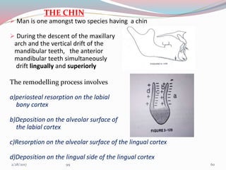
THE CHIN
 Man is one amongst two species having a chin
 During the descent of the maxillary
arch and the vertical drift of the
mandibular teeth, the anterior
mandibular teeth simultaneously
drift lingually and superiorly
The remodelling process involves
a)periosteal resorption on the labial
bony cortex
b)Deposition on the alveolar surface of
the labial cortex
c)Resorption on the alveolar surface of the lingual cortex
d)Deposition on the lingual side of the lingual cortex
603/1/2017 99
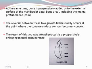
 At the same time, bone is progressively added onto the external
surface of the mandibular basal bone area , including the mental
protuberance (chin).
 The reversal between these two growth fields usually occurs at
the point where the concave surface contour becomes convex.
 The result of this two way growth process is a progressively
enlarging mental protuberance
613/1/2017 99
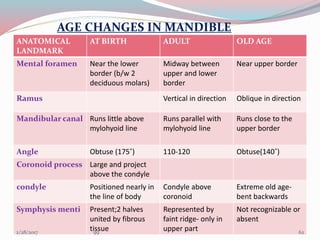
AGE CHANGES IN MANDIBLE
ANATOMICAL
LANDMARK
AT BIRTH ADULT OLD AGE
Mental foramen Near the lower
border (b/w 2
deciduous molars)
Midway between
upper and lower
border
Near upper border
Ramus Vertical in direction Oblique in direction
Mandibular canal Runs little above
mylohyoid line
Runs parallel with
mylohyoid line
Runs close to the
upper border
Angle Obtuse (175˚) 110-120 Obtuse(140˚)
Coronoid process Large and project
above the condyle
condyle Positioned nearly in
the line of body
Condyle above
coronoid
Extreme old age-
bent backwards
Symphysis menti Present;2 halves
united by fibrous
tissue
Represented by
faint ridge- only in
upper part
Not recognizable or
absent
623/1/2017 99
THEORIES OF MANDIBULAR GROWTH
GENETIC THEORY:-
This theory states that all growth is compelled by genetic
influence ie: genetic encoding of mandible determines its
growth.
CARTILAGENOUS THEORY
 This theory states that the cartilage is the primary
determinant of skeletal growth while bone responds
secondarily & passively.
 According to this theory, the condyle by means of
endochondral ossification deposits bone, which tends to
grow the mandible.
633/1/2017 99
ENLOW’S EXPANDING ‘V’ PRINCIPLE
 This theory states that many facial bones or a part of the bone
follows a ‘v’ pattern of enlargement.
Deposition is in the inner surface of of ‘v’ .
Resorption is seen along the outer surface of ‘v’.
CORONOID PROCESS: Deposition –lingualsurface,
Resorption-buccal
CONDYLE PROCESS: Deposition-ant. & post. Margins,
Resorption-buccal & lingual surfaces
643/1/2017 99
ENLOW’S COUNTERPART PRINCIPLE
This principle states that growth of any given facial or cranial
part relates specifically to other structural & geometric
counterpart in the face & cranium
653/1/2017 99
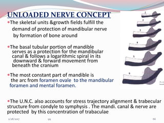
UNLOADED NERVE CONCEPT
The skeletal units &growth fields fulfill the
demand of protection of mandibular nerve
by formation of bone around
The basal tubular portion of mandible
serves as a protection for the mandibular
canal & follows a logarithmic spiral in its
downward & forward movement from
beneath the cranium
The most constant part of mandible is
the arc from foramen ovale to the mandibular
foramen and mental foramen.
The U.N.C. also accounts for stress trajectory alignment & trabecular
structure from condyle to symphysis . The mandi. canal & nerve are
protected by this concentration of trabaculae
663/1/2017 99
SERVO SYSTEM THEORY
 Control of primary cartilages takes a cybernetic form of ‘command’ whereas
control of secondary cartilage like condyle is comprised of both direct effect
of cell multiplication and also indirect effects
Theories of craniofacial growth in the postgenomic area ;semin ortho 2005 ,11:172-83
MUSCLE
FUNCTIONS
actuator
ANTERIOR GROWTH
OF MIDFACE
Reference input
OCCLUSAL
DEVIATION
comparator
Regulation of
inputs-CNS
controller
TRIGGERS –
PROPRIOCEPTIVE
RECEPTORS(MUSCLES &
PERIODONTIUM
CONDYLAR
GROWTH
HORMONAL
FACTORS-
Command
673/1/2017 99
GNOMONIC GROWTH & LOGARITHMIC SPIRAL
A mathematical model was proposed by Moss that describes
mandibular growth along a logarithmic spiral
This was based on D’Arcy Thompsons study on sea-shells
( Nautilus)
Chambered Nautilus
The characteristics of its growth are
 Original shape remains constant,
with increase in size.
 Gnomonic growth can be described
by a curve called as equiangular or
logarithmic spiral
683/1/2017 99
“Gnomon” – that portion or increment
which when added doesn’t alter the shape
but only produces an increase in size
Equiangular or Logarithmic spiral – The growth of the nautilus
follows a particular spiral.
 The important feature of the spiral is
movement of point away from pole along
the radius vector with velocity increasing
with distance from pole
 The angles formed with pole are equal
693/1/2017 99
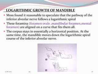
LOGARITHMIC GROWTH OF MANDIBLE
 Moss found it reasonable to speculate that the pathway of the
inferior alveolar nerve follows a logarithmic spiral
 These foramina (foramen ovale ,mandibular foramen,mental
foramen) are aligned on a curve that fits them all.
 The corpus stays in essentially a horizontal position. At the
same time, the mandible moves down the logarithmic spiral
course of the inferior alveolar nerve.
703/1/2017 99
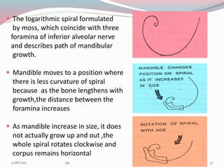
 The logarithmic spiral formulated
by moss, which coincide with three
foramina of inferior alveolar nerve
and describes path of mandibular
growth.
 Mandible moves to a position where
there is less curvature of spiral
because as the bone lengthens with
growth,the distance between the
foramina increases
 As mandible increase in size, it does
not actually grow up and out ,the
whole spiral rotates clockwise and
corpus remains horizontal
713/1/2017 99
FUNCTIONAL MATRIX THEORY
According to the functional Matrix theory Logarithmic growth is
related to active and passive processes:
Active transformative skeletal growth process occurring at the
level of the skeletal units (histologically discernible).
Passive translative - primary expansive growth of their capsular
functional matrices by an alteration in their spatial position.
As the orofacial capsule expands the embedded mandible is
passively lowered in space. The capsular matrix expansion is
not haphazard but involves postnatal rotation of the inferior
alveolar nerve about an axis passing through fixed axis
(foramen ovale).
723/1/2017 99
 This suggests that anterior positioning of the mental foramen
must be a passive growth event. But the mandibular foramen
actively grows up back and out.
 Hence the distance between the mandibular and mental foramen
increases, as does the distance between Ovale and Mandibular
foramina. All these increases however are allometric.
 The position, angulation, resting lengths and tensions of the
masseter and medial pterygoid muscles tend to be altered by the
passive expansion of the orofacial capsule as a consequence of the
rotation of the logarithmic curve.
- Neuromuscular adaptation
- Neurotrophic regulation
733/1/2017 99
passive growth of oro-facial capsule
primary alterations in resting length
contractile and passive
tension, and /or angulation of
the related muscles
change in the growth of the skeletal units
alter the muscle activity vectors
Active mandibular skeletal growth
CNS
Membrane
conduction
Neuromuscular
Adaptation
743/1/2017 99
PROBLEMS OF MANDIBULAR GROWTH AND THEIR
ORTHODONTIC SIGNIFICANCE
HYPOGNATHISM
Agnathia - mandible may be grossly deficient or absent which
reflects deficiency of neural crest cell tissue in lower part of the
face.
First arch and second arch syndrome – Aplasia of mandible
and hyoid bone.
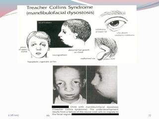
Micrognathia - a diminutive mandible, occurs in
Pierre Robin’s syndrome
Cat cry syndrome
Mandibulofacial dysostosis
Progeria
Down’s syndrome
Oculo-auriculo vertebral syndrome
Turner’s syndrome
753/1/2017 99
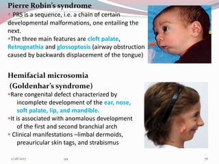
Pierre Robin’s syndrome
 PRS is a sequence, i.e. a chain of certain
developmental malformations, one entailing the
next.
The three main features are cleft palate,
Retrognathia and glossoptosis (airway obstruction
caused by backwards displacement of the tongue)
Hemifacial microsomia
(Goldenhar’s syndrome)
Rare congenital defect characterized by
incomplete development of the ear, nose,
soft palate, lip, and mandible.
It is associated with anomalous development
of the first and second branchial arch
 Clinical manifestations –limbal dermoids,
preauricular skin tags, and strabismus
763/1/2017 99
773/1/2017 99
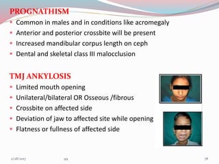
PROGNATHISM
 Common in males and in conditions like acromegaly
 Anterior and posterior crossbite will be present
 Increased mandibular corpus length on ceph
 Dental and skeletal class III malocclusion
TMJ ANKYLOSIS
 Limited mouth opening
 Unilateral/bilateral OR Osseous /fibrous
 Crossbite on affected side
 Deviation of jaw to affected site while opening
 Flatness or fullness of affected side
783/1/2017 99
CONDYLAR HYPERTROPHY
 Mostly due to genetic or hormonal causes
 Common in males
 Usually expressed in late teen age when the growth of mandible
continues at condyle
 More likely to be a high angle case
 Unilateral-chin divergent on side opposite to hypertrophy
facial assymetry
buccal crossbite on unaffected side
793/1/2017 99
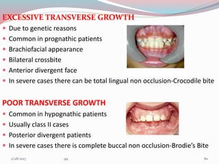
EXCESSIVE TRANSVERSE GROWTH
 Due to genetic reasons
 Common in prognathic patients
 Brachiofacial appearance
 Bilateral crossbite
 Anterior divergent face
 In severe cases there can be total lingual non occlusion-Crocodile bite
POOR TRANSVERSE GROWTH
 Common in hypognathic patients
 Usually class II cases
 Posterior divergent patients
 In severe cases there is complete buccal non occlusion-Brodie’s Bite
803/1/2017 99
PROBLEMS OF RAMAL GROWTH
 EXCESSIVE VERTICAL RAMAL GROWTH
Brachiofacial patients
low angle cases
anterior deep bite
 POOR VERTICAL RAMAL GROWTH
dolichofacial patients
high angle cases
anterior open bite
 EXCESSIVE HORIZONTAL RAMAL GROWTH
More broad oropharynx
 POOR HORIZONTAL RAMAL GROWTH
Narrow oropharynx
Chances of airway embarassment
813/1/2017 99
PROBLEMS OF CHIN GROWTH
PROMINENT CHIN
Common in males
Due to late gonial deposition
Excessive mental bone resorption
Can be treated with genioplasty in adults
DEFECTS DUE TO FAILURE OF FUSION OF PROCESSES
MANDIBULAR CLEFT; Rare condition due
to persistence of furrow between 2
mandibular processes
MICROSTOMIA/MACROSTOMIA;Determined by fusion of maxillary and
mandibular process at their lateral extent 823/1/2017 99
SIGNIFICANCE
 Timely identification of growth disturbances helps in
interception of developing malocclusions and other
orthodontic and esthetic facial problems
 Knowing the timing of development of different facial
structures gives you idea about the long term facial
apppearence of the patient
 Timely diagnosis of growth problems gives you a chance to
treat the problem with functional appliances
833/1/2017 99
Role of functional appliances in the growth of mandible
MANDIBULAR DEFICIENCY
A skeletal Class II relationship could be either due to a small
mandible or a normal mandible in posterior position.
 One possibility of treatment is to restrain the growth of maxilla
with extra oral force and let the mandible continue to grow
more or less normally
 Enhancement of mandibular skeletal growth and this is done
with the help of functional appliance which hold the mandible
forward from its retruded position and enhances growth.
843/1/2017 99
 For most mandibular deficient patients: a standard bionator or
activator appliance is used as it is a simple, durable and readily
acceptable appliance.
 If transverse expansion is needed ,buccal loops attached to bionator
or buccal shields of Frankel appliance shields the cirumoral
musculature away and thus helps in transverse expansion
 Twin block appliance comprising of upper and lower acrylic blocks
which works together and helps in positioning lower jaw forward
 The Herbst appliance ,a fixed functional appliance can also be used
to correct class II malocclusion due to retrognathic mandible
 Mandibular anterior repositioning appliance , Forsus appliance,
Cemented twin block can also be used 853/1/2017 99
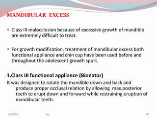
MANDIBULAR EXCESS
 Class III malocclusion because of excessive growth of mandible
are extremely difficult to treat.
 For growth modification, treatment of mandibular excess both
functional appliance and chin cup have been used before and
throughout the adolescent growth spurt.
1.Class III functional appliance (Bionator)
It was designed to rotate the mandible down and back and
produce proper occlusal relation by allowing max.posterior
teeth to erupt down and forward while restraining eruption of
mandibular teeth.
863/1/2017 99
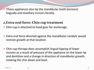
 These appliances also tip the mandibular teeth (incisors)
lingually and maxillary incisors facially.
2.Extra oral force: Chin cup treatment
 Chin cup is attached to head gear for anchorage.
 Extra oral force directed against the mandibular condyle would
restrain growth at that location.
 Chin cup therapy does accomplish lingual tipping of lower
incisors as a result of pressure of the appliance on the lower lip
and dentition and a change in direction of mandibular growth,
rotating the chin down and back.
873/1/2017 99
A study was conducted to evaluate the dental and skeletal effects of chin
cup using two different force magnitudes
 Fifty growing class III patients were divided into three groups.
Patients in group 1 (n  =  20) - 600 g of force per side.
Patients in group 2 (n  =  20) - 300 g of force per side.
Group 3 (n  =  10) no treatment was performed
Lateral Cephalograms were traced and analyzed before treatment and after one year
They concluded that;
 The use of a chin cup significantly improved the mandibular and maxillary
relationship, but with only minor skeletal effects.
 Reduced ramus height and increased the anterior facial height, mandibular
plane angle, and retroclination of the mandibular incisors.
 Use of 600g force had a more pronounced effect in the reduction of ramus
height.
Chin cup effects using two different force magnitudes in the management of Class III
malocclusions,
Yasser L. Abdelnaby and Essam A. Nassar THE ANGLE ORTHODONTIST;SEP.2010 :VOL.8 ,ISS. 5883/1/2017 99
ENVELOPE OF DISCREPENCY (william R Proffit ,Ackerman J. L)
It has three envelopes .The perimeter of each envelope gives the
maximum range of movements possible by different methods
INNER ENVELOPE- Only orthodontic treatment
MIDDLE ENVELOPE- Orthodontic &growth modification
OUTERMOST ENVELOPE- Orthodontic & surgical treatment
893/1/2017 99
10
6
5
5
903/1/2017 99
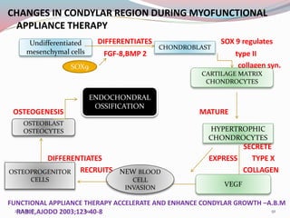
CHANGES IN CONDYLAR REGION DURING MYOFUNCTIONAL
APPLIANCE THERAPY
DIFFERENTIATES SOX 9 regulates
FGF-8,BMP 2 type II
collagen syn.
OSTEOGENESIS MATURE
SECRETE
DIFFERENTIATES EXPRESS TYPE X
RECRUITS COLLAGEN
FUNCTIONAL APPLIANCE THERAPY ACCELERATE AND ENHANCE CONDYLAR GROWTH –A.B.M
RABIE,AJODO 2003;123-40-8
Undifferentiated
mesenchymal cells
SOX9
CHONDROBLAST
CARTILAGE MATRIX
CHONDROCYTES
HYPERTROPHIC
CHONDROCYTES
VEGF
NEW BLOOD
CELL
INVASION
OSTEOPROGENITOR
CELLS
OSTEOBLAST
OSTEOCYTES
ENDOCHONDRAL
OSSIFICATION
913/1/2017 99
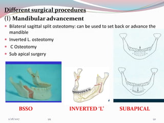
Different surgical procedures
(I) Mandibular advancement
 Bilateral sagittal split osteotomy: can be used to set back or advance the
mandible
 Inverted L. osteotomy
 C Osteotomy
 Sub apical surgery
BSSO INVERTED ‘L’ SUBAPICAL
923/1/2017 99
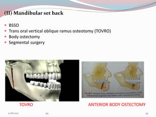
(II) Mandibular set back
 BSSO
 Trans oral vertical oblique ramus osteotomy (TOVRO)
 Body ostectomy
 Segmental surgery
TOVRO ANTERIOR BODY OSTECTOMY
933/1/2017 99
Correction of Mandibular Retrognathia and Laterognathia by
Distraction Osteogenesis: Follow up of 5 cases-EUR J DENT2009
Oct; 3(4): 335–342.
 The procedure was carried out in 5 subjects (3 males and 2 females)aged
between 14 years and 27 years.3 Patients had undergone bilateral
distraction osteogenesis and remaining 2 ,unilateral distrctn.osteogenesis
 In patients treated with bilateral mandibular distraction, it was observed
that the ANB angle decreased by a mean of 5°, the mandibular corpus
length increased by a mean of 14.5 mm and the overjet decreased by a
mean of 12.2 mm after treatment.
 In patients treated with unilateral mandibular distraction, a mean of 3.5°
reduction was achieved in ANB angle, the mandibular corpus length
increased by a mean of 5.5 mm and a mean of 7 mm correction was
achieved in relation to craniofacial midline with treatment.
 It can be concluded that distraction of the deformed mandible is a
feasible and effective technique for treating mandibular retrognathia and
laterognathia and that long term relapse is within acceptable limits. 943/1/2017 99
Mandibular Growth, Remodeling, and Maturation During Infancy
and Early Childhood(Yi-Ping Liu, Rolf G. Behrents, and Peter H.
Buschang) The Angle Orthodontist-vol.80 Jan 2010
 In a study lateral cephalograms of 24 females and 24 males, taken
between birth and 5 years of age, as well as early adulthood, were
traced and digitized. Five measurements and nine landmarks were used
to characterize mandibular growth, remodeling, and degree of adult
maturity.
Ramus height (Co-Go),Overall length (Co-Gn),Corpus length (Go-Gn),Condylion
angle (Go-Co-Me),Gonial angle (Co-Go-Me)
They concluded that
1. Mandibular size increased 18.2 mm to 34.7 mm between 0.4 and
5.0 years of age.
2. Males displayed greater growth increases for ramus height (Co-Go)
than for corpus length (Go-Gn), and females showed similar changes
3. Gonial angle decreased 2.8° and 2.0° in males and females,
respectively. 953/1/2017 99
Regulation of the Response of the Adult Rat Condyle to
Intermaxillary Asymmetric Force by the RANKL-OPG System(Yue
Xua; Tuojiang Wub; Yangxi Chenc; Zhiguang Zhangd) The Angle
Orthod. 2009;79:646–651.
 The mandibular rami of 160 Sprague-Dawley rats (3 months old) were
subjected to unilateral traction in the anterior-superior direction using
an elastic force. (120 & 40 g ,then traction removed after28 days.
 The expression of RANKL and OPG in the subchondral bone of the
condyles was analyzed by semiquantitative immunohistochemistry.
Results:
- Different force levels induced similar changes in the expression of the
OPG protein by28 days.
-The effect of a 120-g elastic force on the expression of RANKL was
stronger than that of a 40-g force. Because of the asynchrony of RANKL
responses to external forces of different values, the values of
RANKL/OPG ratio showed characteristic variation
963/1/2017 99
CONCLUSION
Bone growth in mandible is a remodelling process
represented by apposition and resorption.
Knowledge of general facial growth provides a
background to the understanding of the etiology
and development of of malocclusion, such an
understanding is in turn an important part of
diagnosis and treatment planning.
973/1/2017 99
REFERENCES
1.HANDBOOK OF FACIAL GROWTH,SECOND EDTN-ROBERT E MOYERS
2.THE HUMAN FACE-DONALD H ENLOW
3.HUMAN EMBRYOLOGY,EIGHTH EDITION-INDERBIR SINGH
4.ORTHODONTIC CURRENT PRINCIPLES & TECHNIQUE,FIFTH EDITION-
T.MGRABER
5.CONTEMPORARY ORTHODONTICS,FIFTH EDITION-WILLIAM R PROFITT
6.DENTOFACIAL ORTHOPEDICS WITH FUNCTIONAL APPLIANCES-
GRABER,RAKOSI,PETROVIC
7.CRANIOFACIAL DEVELOPMENT-GEOFFREY H SPERBER
8.PRENATAL DEVELOPMENT OF HUMAN MANDIBLE.THE ANATOMICAL
RECORDS 263:314-325(2001)
9.GROWTH OF MANDIBLE DURING PUBESCENCE-ROCHE,LEWIS AO OCT
1982
10.AGE RELATED DIFFERENCE IN RAMUS GROWTH-ENLOW,HANS AJO
DEC.1995
.
983/1/2017 99
11. VARIATION IN GROWTH PATTERN OF HUMAN MANDIBLE;A
LONGITUDINAL RADIOGRAPHIC STUDY BY IMPLANT METHOD-
ARNE BJORK ;J DENT RES 1963
12.CORRECTION OF MANDIBULAR RETROGNATHIAAND
LATEROGNATHIA BY DISTRACTION OSTEOGENESIS-EUR J DENT
2009 OCT:3(4);335-342
13. MANDIBULAR GROWTH,REMODELLING AND MATURATION
DURING INFANCYAND CHILDHOOD-ROLF G BEHRENTS,PETER H
BUSCHANG-AO JAN.2010
14.THEORIES OF CRANIOFACIAL GROWTH IN POSTGENOMIC
ERA;DAVID S CARLSON: SEM ORTHO ,2005 11:172–183
15. REGULATION OF THE RESPONSE OF THE ADULT RAT CONDYLE TO
INTERMAXILLARY ASYMMETRIC FORCE BY THE RANKL-OPG
SYSTEM-YUE XUA,TUOIJANG WUB.THE ANGLE ORTHODONTIST
2009;79,646-651
16.FUNCTIONALAPPLIANCE THERAPY ACCELERATE AND ENHANCE
CONDYLAR GROWTH –A.B.M RABIE,AJODO 2003;123-40-8
993/1/2017 99
More Related Content

growth and development of mandible

  • 1. Dr SHILPA JOY V S Dental College 13/1/2017 99
  • 2. CONTENTS  Introduction  Anatomy of mandible  Evolution of mandible  Mandibular growth mechanisms-Brief history  Prenatal growth of mandible  Postnatal development  Growth progression-mechanism & site  Age changes in mandible  Theories of mandibular growth  Problems of mandibular growth and orthodontic significance  Conclusion  References 23/1/2017 99
  • 3. INTRODUCTION The human mandible has no one design for life.Rather it adapts and remodels through the seven stages of life,from the slim arbiter of things to come in the infant,through a powerful dentate machine and even weapon in the full flesh of maturity ,to the pencil thin,porcelain like problem that we struggle to repair in the adversity of old age –D.E POSWILLO It is attached only by ligaments and muscles to immovable bones of the skull. The temporomandibular or ginglymo-diarthroidal joints are the only visible movable articulations in the head. The rest of the bones of the skull move in union when the head is moved as a whole. 33/1/2017 99
  • 4. DEFINITIONS RELATED TO GROWTH  Moss: change in morphological parameters which is measurable.  Moyers: Quantitative aspect of biological development per unit of time.  Todd: An increase in size.  Krogman: increase in size change in proportions and progressive complexity  J.S. Huxley: The self multiplications of living substance  Meridith:entire series of sequential anatomic and Physiologic changes taking place from beginning of pre natal life to serenity DEVELOPMENT  Todd:progress towards maturity  Moyers:All naturally occuring unidirectional changes in the life of an individual from its existence as a single cell to its elaboration as a multifunctional unit terminating in death 43/1/2017 99
  • 7. Attachments and relations of mandible  Oblique line – buccinator muscle  Oblique line below mental foramen – depressor labii inferious, depressor anguli oris  Lateral side of ramus- Masseter muscle  Lower border- Platysma  Midsurface of coronoid- Temporalis  Incisive fossa- Mentalis 73/1/2017 99
  • 8.  Postero superior part of lateral surface – Parotid gland  Lingula – sphenomandibular ligament  Pterygoid fossa - lateral pterygoid muscle  Mylohyoid line- Mylohyoid muscle  Posterior end of mylohyoid line- superior constrictor muscle  Digastric fossa- Anterior belly of digastric  Genial tubercle -Genioglossus & Geniohyoid 83/1/2017 99
  • 9. EVOLUTION OF MANDIBLE  The agnatha, the earliest type of vertebrate, had its mouth opening on the ventral side anteriorly along the vertebral axis. They did not have jaws  The Placoderms, had 7 arches.The first arch was lost.Their new first arch became the Mandibular arch that formed the jaws.The upper half of mand.arch became palatoquadrate cartilage and lower half became the mandibular or meckels cartilage  In Elasmobrancs,the jaws were formed by mandibular arch (arch 1) & hyoid arch(arch 2) The upper half of hyoid arch became hyomandibular ligament and lower half became hyoid cartilage 93/1/2017 99
  • 10. In amphibians the hyomandibular ligament became the stapes .They had a dentary bone in the anterior end of the original cartilaginous jaw. At its posterior extremity it articulated with the quadrate bone In reptiles like early synapsid ,that gave rise to mammals ,the jaw joint is formed by the articular(lower) & quadrate(upper) bones.The joint was a simple hinge at the posterior of jaw In mid and late synapsid reptiles,the dentary bone (lower jaw) increase in size as muscle and bite force increased,but force on the joint decreased as the muscle insertion point shifted to allow greater jaw mobility The articular and quadrate bones at the jaw joint became smaller and was loosely attached with the dentary 103/1/2017 99
  • 11.  The coronoid process of dentary bone formed to accommodate these changing forces  Ultimately in mammals,the jaw joint shifted from a articular-quadrate joint to a dentary-squamosal joint.the condylar process formed to create a new articulating surface 113/1/2017 99
  • 12. MANDIBULAR GROWTH PATTERNS- A HISTORY • HUNTER (1771) compared a series of dried mandibles and concluded that in order to attain space for permanent molar teeth the mandible must grow by posterior apposition of ramus accompanied by anterior ramus resorption. • HUMPHRY (1866) studied growth of mandible by inserting metal rings in the anterior and posterior margins of mandibular ramus in growing pig.Rings placed on posterior border became more deeply embedded but rings placed on anterior surface were released • BRASH (1924) fed pigs the madder plant root(alizarin ) which labeled appositional growth  WEINMAN AND SICHER (1940) with the help of longitudinal cephalometrics and evidence from experiments of animals, focused attention on the mandibular condyle as a major factor in growth of the mandible. A STUDY OF POSTNATAL GROWTH OF HUMAN MANDIBLE-DONALD H ENLOW,DAVID B HARRIS AJODO,JAN 1964 123/1/2017 99
  • 13.  BRODIE believed that superior and posterior growth of the condyle along with apposition of the posterior border of the ramus and alveolar border resulted in development of mandible.  RICKETTS(1950)by superimpositions on lower border of the mandible showed that the condyle followed a superior and posterior course. He also noted that the mandibular growth was not same and that the relationship of the mandibular plane to the Frankfort Horizontal plane was changing about one degrees every 3 yrs in a typical facial pattern.  MOSS(1960) envisioned the growth of the mandible as a logarithmic spiral constructed via the path of the mandibular nerve. 133/1/2017 99
  • 14.  BJORK (1963 )conducted a study with tantalum implants and suggested that 1. Growth in length of the mandible occurs at the condyles. 2. The anterior aspect of chin is extremely stable 3. The thickening of the symphysis takes place by appostion on its posterior surface and lower border which contributes to increase in height of symphysis. 4. At the region of the condyles there is upward and forward curving growth. 5. The mandibular canal is not remodeled and the trabaculae related to the canal are stationary. Hence the curvature of the mandibular canal generally reflects the earlier shape of the mandible VARIATION IN GROWTH PATTERN OF HUMAN MANDIBLE;A LONGITUDINAL RADIOGRAPHIC STUDY BY IMPLANT METHOD-ARNE BJORK ;J DENT RES 1963 . 143/1/2017 99
  • 15. Growth & development of an individual can be divided in to Pre-natal Post-natal Period of Ovum 1-14th day Period of embryo 14th – 56th day Period of Fetus 56th – 270th day 153/1/2017 99
  • 16. PRENATAL GROWTH PERIOD OF OVUM Fertilization-Ampulla of uterine tube zygote mitosis Cluster of cells(Blastomere) mitosis Morula(16 cell structure) Blastocyst 163/1/2017 99
  • 17. BLASTOCYST Some fluid passes into morula from uterine wall seperating the inner cell mass (embryoblast) and outer cell mass (trophoblast) Trophoblast cells become flattened and embryoblast cells get attached to one side Now it is called blastocyst and cavity is called blastocoele The site of attachment of inner cell mass is called embryonic or animal pole and opposite site abembryonic pole MORULA 173/1/2017 99
  • 18. PERIOD OF EMBRYO PRESOMITE PERIOD(8-20th day)  Trophoblastic layer differentiate into synctiotrophoblast and cytotrophoblast layers SYNCTIOTROPHOBLAST- Outer cells that invades endometrium and its vessels to establish maternal blood circulation to developing embryo-UTEROPLACENTAL CIRCULATION PRESOMITE SOMITE POSTSOMITE 183/1/2017 99
  • 19. INNER CELL MASS Differentiation BILAMINAR DISC BLASTOCYSTIC CAVITY is now called as primitive yolk sac AMNIOTIC CAVITY develops between epiblast and cytotrophoblast HYPOBLAST Squamous /cuboidal EPIBLAST Columnar cells 193/1/2017 99
  • 20.  Extra embryonic mesoderm(EEM) formed of loose connective tissue , differentiate between developing embryo and cytotrophoblast  Chorionic cavity is formed by fusion of number of lacunae that develop in EEM  Expansion of chorionic cavity reduces size of primitive yolk sac,forming secondary yolk sac, occurs by end of second week  3rd week- Gastrulation occurs Bilaminar disc-Trilaminar disc 203/1/2017 99
  • 21.  Cells of primitive streak grow cranially to reach the prochordal plate to form notochord which is a solid cylinder of cells,axial skeleton of fetus forms around notochord NEURAL TUBE FORMATION Ectoderm above notochord thickens Neural plate Midline of neural plate deepens Neural groove 213/1/2017 99
  • 22. Elevated margins on either side-Neural folds Neural folds grow towards each other Fuse to form Neural tube- CNS Edges of neural tube on either side-neural crests Anterior end of neural tube-fore,mid,hind brain Certain elevations called rhombomeres in area of hind brain-cells that proliferate from neural crests 223/1/2017 99
  • 23. NEURAL CREST CELLS Forms from neuro ectoderm Migrate & differentiate extensively with in the developing embryo Spinal & cranial sensory ganglia, Sympathetic neurons, Schwann cells, pigment cells & meninges Most of the connective tissue of the head is formed Migration is essential for development of teeth & face All the tissues of teeth (except enamel) & its supporting apparatus are derived directly from these cells 233/1/2017 99
  • 24. SOMITE PERIOD(21ST -31ST day of IUL)  Rapid growth of cranial end of embryo,caudal end lags behind- CEPHALOCAUDAL GRADIENT OF GROWTH Head-1/2 of total embryonic disk length BRANCHIAL /PHARYNGEAL ARCHES  In specific areas,the migrating and rapidly proliferating ectomesenchyme cells develops elevation between ectoderm and endoderm  4th week of IUL Elevations seen in ventral foregut 5th arch perishes Formation of 6 pharyngeal arches (bilaterally) Finally 5 arches remain 243/1/2017 99
  • 25. Separated externally by small clefts called branchial grooves (Ectodermal clefts)-4 in number On the inner aspect of pharyngeal wall are corresponding small depressions called pharyngeal pouches-5 in number In aquatic vertebrates both branchial grooves & pharyngeal pouches fuse to form gill slits 253/1/2017 99
  • 27. DEVELOPMENT OF MANDIBLE Develops from the mandibular process of 1st branchial arch The cartilage of the 1st arch (Meckle’s cartilage) forms lower jaw in the primitive vertebrates In human beings Meckel’s cartilage has close positional relationship to the developing mandible but makes no contribution to it The mandibular nerve has close relationship to the Meckel’s cartilage, beginning 2/3 of the way along the length of cartilage At this point mandibular nerve divides in to lingual and inferior alveolar branches 273/1/2017 99
  • 28. At around 36-38 days of IUL there is ectomesenchymal condensation Some mesenchymal cells enlarges,acquire a basophilic cytoplasm and form osteoblasts Osteoblast secrete a gelatinous matrix called osteoid and results in ossification of osteogenic membrane The resulting intramembranous bone lies lateral to meckels cartilage of mandibular arch 283/1/2017 99
  • 29. In sixth wk ,a single ossification centre for each half arises in the bifurcation of inferior alveolar nerve into mental and incisive 7th wk-bone begin to develop lateral to meckels cartilage and continues until the postr aspect is covered with bone Between 8th & 12th wk ,mandibular growth accelerate ,as a result mandibular length increases 293/1/2017 99
  • 30. Ossification stops at apoint,which later become lingula,the remaining part of meckels cartilage continues to form sphenomandibular ligament &spinous process of sphenoid Secondary accessory cartilage appears between 10th &14th wk to form head of condyle,part of coronoid process & mental protuberance 303/1/2017 99
  • 31. FATE OF MECKELS CARTILAGE Posterior extremity forms malleus, incus & sphenomandibular ligament Most of the cartilage is absorbed except for some portion in midline which may cause endochondral ossification FETAL PERIOD Endochondral bone formation seen only in 3 areas • Condylar process • Coronoid process • Mental region 313/1/2017 99
  • 32. Condylar process  About 5th week of I.U.L. area of mesenchymal condensation above the ventral part of developing mandible  About 10th wk develops into cone shaped cartilage  By 14th week starts ossification  By 4 months migrates inferiorly and fuses with ramus  4th month onwards replaced by bone but proximal end persists into adulthood acting as Growth cartilage & Articular cartilage  Condylar head separated from temporal bone by thin disc of connective tissue – future articular disc 323/1/2017 99
  • 33. Coronoid process  By 10th to 14th week of I.U.L. secondary cartilages seen in region of coronoid  This cartilage becomes incorporated into expanding intramembranous bone of ramus and disappears before birth Mental region  Secondary cartilages seen on both sides -- ossify by 7th wk I.U.L.  They ossify to form mental ossicles in fibrous tissue of symphysis and later on gets incorporated into it. 333/1/2017 99
  • 34. POST NATAL DEVELOPMENT MANDIBLE AT BIRTH MANDIBULAR GROWTH-FIRST YEAR GROWTH PROGRESSION AFTER FIRST YEAR- MECHANISM & SITE 343/1/2017 99
  • 35. MANDIBLE AT BIRTH  2 rami of mandible are quite short,wide and the condyles are poorly developed  The alveolar process not yet formed  The body of mandible is like a shell of bone with tooth follicles and developing crowns of teeth which are covered by occlusal gum pads  The angle of the mandible is about 175 degree with condyle nearly in line with the body  Initial separation of the two mandibular halves by fibrocartilage and connective tissue which is eliminated gradually as ossification occurs between 4th month to 1st year. 353/1/2017 99
  • 36. MANDIBULAR GROWTH DURING FIRST YEAR Appositional growth especially active at  Alveolar border  Distal and superior surface of ramus  Condyle  Lower border of mandible  Lateral surface of mandible  After first year growth becomes more selective,condyle shows considerable activities , mandible moves and grows forward and downward 363/1/2017 99
  • 37. GROWTH PROGRESSION-MECHANISM & SITE MANDIBULAR CONDYLE  It is a major site of growth Historically, the condyle has been regarded as a kind of cornucopia from which the whole mandible itself pours forth.  During mandibular growth ,the condyle functions as regional field of growth that provides an adaptation for its own localized growth circumstances 373/1/2017 99
  • 38.  The condylar cartilage is a secondary type of cartilage  Its real contribution is to provide regional adaptive growth  Main functional role of condyle is (1) provides a pressure tolerant articular contact (2) it makes possible a multidimensional growth capacity in response to ever-changing, developmental conditions and variations.  The condylar growth mechanism itself is a clear-cut process. Cartilage is a special non-vascular tissue and is involved because variable levels of compression occur at its articular contact with temporal bone 383/1/2017 99
  • 39.  An endochondral growth mechanism is required ,because the condyle grow in a direction towards its articulation in the face of direct pressure  Intramembranous type couldn’t operate because the periosteal mode of osteogenesis is not pressure adapted  In Figure the endochondral bone tissue (b) , formed in association with the condylar cartilage (a) is laid down only in medullary portion  The enclosing bony cortices (c) are produced by periosteal- endosteal osteogenic activity 393/1/2017 99
  • 40. HISTOLOGICALLY Articular zone Resting zone Proliferative zone Hypertrophic zone Erosive zone • In secondary cartilage like condyles,the prechondoblast are not surrounded by cartilaginous matrix ,thus they are exposed to environment and are moldable to external influences •Codylar cellular arrangement is multidirectional unlike primary cartilage where its arranged in rows 403/1/2017 99
  • 41.  Anterior margin of condylar neck is depository, this surface is part of sigmoid notch  Posterior edge which grades into posterior border of ramus is also depository The lingual and buccal sides of neck characteristically have a resorptive surface. This is because condyle is quite broad and neck is narrow 413/1/2017 99
  • 42.  The neck is progressively relocated into areas previously held by the much wider condyle,and it’s sequentially derived from the condyle as condyle moves in superoposterior course  Explained another way, the endosteal surfaceof the neck actually faces the growth direction; the periosteal side points away from the course of growth.  This is another example of the V principle, with the V-shaped cone of the condylar neck growing toward its wide end. 423/1/2017 99
  • 43. What is the physical force that produces the forward and downward primary displacement of mandible ??????? For many years it was presumed that  Cartilage is pressure adapted type of tissue & creates a thrust of mandible against its articular bearing surface  proliferation of cartilage towards its contact thereby pushes the whole mandible away from it. But,Bilaterally condyle- lacking mandibles occupy an essentially normal anatomic position and the mandible functions in movements These observations suggested 2 conclusions. 1. Condyles may not play the kingpin role of a “master center”. 2. The whole mandible can become displaced anteriorly and inferiorly into its functional position without a "push" against the basicranium 433/1/2017 99
  • 44.  The current thinking is that condylar cartilage does have a measure of intrinsic genetic programming .  The cartilage cells are coded and geared to divide ,but extracondylar factors are needed to sustain this activity  So overall mandibular length can be clinically increase or decrease depending on class II or class III if this were done during period of active condylar growth Physiologic indicators Extrinsic & intrinsic biomechanical factors 443/1/2017 99
  • 45. CORONOID PROCESS  The coronoid process has propeller like twist, so that its lingual side faces three general directions all at once posteriorly, superiorly and medially.  When bone is added onto the lingual side of the coronoid process , growth thereby precedes superiorly and this part of ramus increases in vertical dimension 453/1/2017 99
  • 46.  These same deposits of bone on the lingual side also bring about a posterior direction of growth movement .  Produces backward movement of two coronoid process even though deposits on the inside (lingual) surface.  These same deposits on the lingual side also bring about medial direction of growth in order to lengthen corpus  Area occupied by anterior part of ramus in mandible 1 becomes relocated and remodeled into posterior part of corpus in mandible 2. 463/1/2017 99
  • 47. CORPUS / BODY OF MANDIBLE  Outer surface-depository & medial surface(inferior aspect)- Resorptive,remodelling is in the form of ‘L’  Depository area -from the superior half of medial surface of corpus to anterior half of medial surface of ramus(below coronoid)  Resorptive area –from inferior half of medial surface of corpus to posterior half of medial surface of ramus (below condyle) 473/1/2017 99
  • 48. RAMUS  At birth the two rami of mandible are quite short, they grow by the process of direct surface apposition and remodeling.  THE PRINCIPLE GROWTH VECTORS ARE IN POSTERIOR & SUPERIOR DIRECTION  Resorption occurs on the anterior surface of ramus while bone deposition occurs on posterior surface.  Bone growth occurs at the mandibular condyle and along the posterior part of ramus to the same extent as anterior part has undergone resorption 483/1/2017 99
  • 49.  The lower part of ramus below the coronoid process also has a twisted contour.  Its buccal side faces posteriorly toward the direction of backward growth and thus characteristically has a depository type of surface.  The opposite lingual side, being away from direction of growth, is resorptive. 493/1/2017 99
  • 50. IMPORTANCE OF RAMUS  It positions the lower arch in occlusion with the upper.  It is continuously adaptive to the multitude of changing craniofacial conditions.  Attach the mastication muscle and must accommodate the increasing mass of masticatory muscle inserted into it.  Bridges the pharyngeal compartment.  The horizontal breadth of ramus determines the anteroposterior positioning of lower arch.  Height of ramus accommodates the vertical dimension and growth of nasal and masticatory components of face.  Remodeling and relocation give space to accommodate erupting permanent molar. 503/1/2017 99
  • 51. RAMUS UPRIGHTING  Greater amounts of bone additions on the inferior part of the posterior border than on the superior part.  A correspondingly greater amount of matching resorption on the anterior border takes place inferiorly than superiorly.  A "remodeling" rotation of ramus alignment thus occurs.  In diagram the pharynx enlarges horizontally from a to a’ .  The ramus enlarges correspondingly from b to b’  Angle c is reduced to c’ to accommodate the vertical increase,which allows for considerable extent of vertical nasomaxillary growth 513/1/2017 99
  • 52.  Vertical lengthening of the ramus continues to take place after horizontal ramus growth slows or ceases  Resorption takes place on the upper part of the posterior border.  A forward growth direction can occur on the anterior border in the upper part of the coronoid process.  A posterior direction of remodeling takes place in the lower part of the posterior border,this result in more upright alignment and longer vertical dimension of ramus without increase in breadth In fig mandible a is superimposed over b remodeling changes outlined above serve simply to alter the ramus angle without increasing its breadth 523/1/2017 99
  • 53. • The growth and remodeling changes of both ramus and middle cranial fossa produces lowering of mandibular arch .This accommodate vertical expansion of nasomaxillary complex . • A vertical imbalance thus occurs ,this ‘opens’ anterior bite,only the first and second molars are in occlusal contact • The amount of upward mandibular tooth drift is much less than the downward drift and displacement of maxillary teeth. • This is one of the several reason why orthodontic purpose, often attack maxillary dentition ,eventhough given malocclusion can be largely based on improper positioning of mandible 533/1/2017 99
  • 54. MANDIBULAR FORAMEN  The mandibular foramen likewise drift backward and upward by deposition on the anterior and resorption on the posterior part of its rim.  The foramen maintains a constant position about midway between the anterior and posterior borders of ramus.  Even when the ramus undergoes marked alterations associated with edentulism ,the foramen usually sustains midway location 543/1/2017 99
  • 55. ANTEGONIAL NOTCH  A single field of surface resorption is present on the inferior edge of mandible at the ramus corpus junction.  This forms the antegonial notch by remodelling from the ramus just behind it as the ramus relocates posteriorly  The size of the notch can be increased whenever a downward rotation of corpus relative to the ramus takes place 553/1/2017 99
  • 56. ANTEGONIAL NOTCH –CLINICAL SIGNIFICANCE  Deep notched subjects have retrusive mandible with shorter corpus, less ramus height and increase gonial angle.  Mandibular growth directions in deep notched patients were more vertically directed as measured by facial axis and the mandibular plane angle.  Deep notched subjects had longer total facial height and longer lower facial height,Smaller saddle angle  Deep notch patients required a longer duration of orthodontic treatment. 563/1/2017 99
  • 57. THE LINGUAL TUBEROSITY  Important structure as it is direct anatomic equivalent of the maxillary tuberosity  Major site of growth for mandible  Effective boundary between basic parts of the mandible ; ramus and corpus.  Grows posteriorly by deposits on the posterior facing surface.  The prominence of tuberosity is increased by presence of large resorptive fields just below it which produces a sizable depression, the lingual fossa. 573/1/2017 99
  • 58.  The combination of resorption in the fossa and deposition on the medial facing surface of tuberoisty itself greatly accentuates the contours of both regions  Deposition on the lingual surface of the ramus just behind the tuberosity produces a medial direction of drift that shifts this part of the ramus into alignment with the axis of corpus. 583/1/2017 99
  • 59. THE RAMUS TO CORPUS REMODELLING CONVERSION  The whole ramus is being relocated in the posterior direction  Bony arch length has been increased and the corpus has been lengthened by -Deposits on the posterior surface of lingual tuberosity and the contiguous lingual side of the ramus. - A resultant lingual shift of this part of ramus to become added to corpus.  The presence of resorption on the anterior border of ramus is usually described as ‘MAKING ROOM FOR THE LAST MOLAR’ 593/1/2017 99
  • 60. THE CHIN  Man is one amongst two species having a chin  During the descent of the maxillary arch and the vertical drift of the mandibular teeth, the anterior mandibular teeth simultaneously drift lingually and superiorly The remodelling process involves a)periosteal resorption on the labial bony cortex b)Deposition on the alveolar surface of the labial cortex c)Resorption on the alveolar surface of the lingual cortex d)Deposition on the lingual side of the lingual cortex 603/1/2017 99
  • 61.  At the same time, bone is progressively added onto the external surface of the mandibular basal bone area , including the mental protuberance (chin).  The reversal between these two growth fields usually occurs at the point where the concave surface contour becomes convex.  The result of this two way growth process is a progressively enlarging mental protuberance 613/1/2017 99
  • 62. AGE CHANGES IN MANDIBLE ANATOMICAL LANDMARK AT BIRTH ADULT OLD AGE Mental foramen Near the lower border (b/w 2 deciduous molars) Midway between upper and lower border Near upper border Ramus Vertical in direction Oblique in direction Mandibular canal Runs little above mylohyoid line Runs parallel with mylohyoid line Runs close to the upper border Angle Obtuse (175˚) 110-120 Obtuse(140˚) Coronoid process Large and project above the condyle condyle Positioned nearly in the line of body Condyle above coronoid Extreme old age- bent backwards Symphysis menti Present;2 halves united by fibrous tissue Represented by faint ridge- only in upper part Not recognizable or absent 623/1/2017 99
  • 63. THEORIES OF MANDIBULAR GROWTH GENETIC THEORY:- This theory states that all growth is compelled by genetic influence ie: genetic encoding of mandible determines its growth. CARTILAGENOUS THEORY  This theory states that the cartilage is the primary determinant of skeletal growth while bone responds secondarily & passively.  According to this theory, the condyle by means of endochondral ossification deposits bone, which tends to grow the mandible. 633/1/2017 99
  • 64. ENLOW’S EXPANDING ‘V’ PRINCIPLE  This theory states that many facial bones or a part of the bone follows a ‘v’ pattern of enlargement. Deposition is in the inner surface of of ‘v’ . Resorption is seen along the outer surface of ‘v’. CORONOID PROCESS: Deposition –lingualsurface, Resorption-buccal CONDYLE PROCESS: Deposition-ant. & post. Margins, Resorption-buccal & lingual surfaces 643/1/2017 99
  • 65. ENLOW’S COUNTERPART PRINCIPLE This principle states that growth of any given facial or cranial part relates specifically to other structural & geometric counterpart in the face & cranium 653/1/2017 99
  • 66. UNLOADED NERVE CONCEPT The skeletal units &growth fields fulfill the demand of protection of mandibular nerve by formation of bone around The basal tubular portion of mandible serves as a protection for the mandibular canal & follows a logarithmic spiral in its downward & forward movement from beneath the cranium The most constant part of mandible is the arc from foramen ovale to the mandibular foramen and mental foramen. The U.N.C. also accounts for stress trajectory alignment & trabecular structure from condyle to symphysis . The mandi. canal & nerve are protected by this concentration of trabaculae 663/1/2017 99
  • 67. SERVO SYSTEM THEORY  Control of primary cartilages takes a cybernetic form of ‘command’ whereas control of secondary cartilage like condyle is comprised of both direct effect of cell multiplication and also indirect effects Theories of craniofacial growth in the postgenomic area ;semin ortho 2005 ,11:172-83 MUSCLE FUNCTIONS actuator ANTERIOR GROWTH OF MIDFACE Reference input OCCLUSAL DEVIATION comparator Regulation of inputs-CNS controller TRIGGERS – PROPRIOCEPTIVE RECEPTORS(MUSCLES & PERIODONTIUM CONDYLAR GROWTH HORMONAL FACTORS- Command 673/1/2017 99
  • 68. GNOMONIC GROWTH & LOGARITHMIC SPIRAL A mathematical model was proposed by Moss that describes mandibular growth along a logarithmic spiral This was based on D’Arcy Thompsons study on sea-shells ( Nautilus) Chambered Nautilus The characteristics of its growth are  Original shape remains constant, with increase in size.  Gnomonic growth can be described by a curve called as equiangular or logarithmic spiral 683/1/2017 99
  • 69. “Gnomon” – that portion or increment which when added doesn’t alter the shape but only produces an increase in size Equiangular or Logarithmic spiral – The growth of the nautilus follows a particular spiral.  The important feature of the spiral is movement of point away from pole along the radius vector with velocity increasing with distance from pole  The angles formed with pole are equal 693/1/2017 99
  • 70. LOGARITHMIC GROWTH OF MANDIBLE  Moss found it reasonable to speculate that the pathway of the inferior alveolar nerve follows a logarithmic spiral  These foramina (foramen ovale ,mandibular foramen,mental foramen) are aligned on a curve that fits them all.  The corpus stays in essentially a horizontal position. At the same time, the mandible moves down the logarithmic spiral course of the inferior alveolar nerve. 703/1/2017 99
  • 71.  The logarithmic spiral formulated by moss, which coincide with three foramina of inferior alveolar nerve and describes path of mandibular growth.  Mandible moves to a position where there is less curvature of spiral because as the bone lengthens with growth,the distance between the foramina increases  As mandible increase in size, it does not actually grow up and out ,the whole spiral rotates clockwise and corpus remains horizontal 713/1/2017 99
  • 72. FUNCTIONAL MATRIX THEORY According to the functional Matrix theory Logarithmic growth is related to active and passive processes: Active transformative skeletal growth process occurring at the level of the skeletal units (histologically discernible). Passive translative - primary expansive growth of their capsular functional matrices by an alteration in their spatial position. As the orofacial capsule expands the embedded mandible is passively lowered in space. The capsular matrix expansion is not haphazard but involves postnatal rotation of the inferior alveolar nerve about an axis passing through fixed axis (foramen ovale). 723/1/2017 99
  • 73.  This suggests that anterior positioning of the mental foramen must be a passive growth event. But the mandibular foramen actively grows up back and out.  Hence the distance between the mandibular and mental foramen increases, as does the distance between Ovale and Mandibular foramina. All these increases however are allometric.  The position, angulation, resting lengths and tensions of the masseter and medial pterygoid muscles tend to be altered by the passive expansion of the orofacial capsule as a consequence of the rotation of the logarithmic curve. - Neuromuscular adaptation - Neurotrophic regulation 733/1/2017 99
  • 74. passive growth of oro-facial capsule primary alterations in resting length contractile and passive tension, and /or angulation of the related muscles change in the growth of the skeletal units alter the muscle activity vectors Active mandibular skeletal growth CNS Membrane conduction Neuromuscular Adaptation 743/1/2017 99
  • 75. PROBLEMS OF MANDIBULAR GROWTH AND THEIR ORTHODONTIC SIGNIFICANCE HYPOGNATHISM Agnathia - mandible may be grossly deficient or absent which reflects deficiency of neural crest cell tissue in lower part of the face. First arch and second arch syndrome – Aplasia of mandible and hyoid bone. Micrognathia - a diminutive mandible, occurs in Pierre Robin’s syndrome Cat cry syndrome Mandibulofacial dysostosis Progeria Down’s syndrome Oculo-auriculo vertebral syndrome Turner’s syndrome 753/1/2017 99
  • 76. Pierre Robin’s syndrome  PRS is a sequence, i.e. a chain of certain developmental malformations, one entailing the next. The three main features are cleft palate, Retrognathia and glossoptosis (airway obstruction caused by backwards displacement of the tongue) Hemifacial microsomia (Goldenhar’s syndrome) Rare congenital defect characterized by incomplete development of the ear, nose, soft palate, lip, and mandible. It is associated with anomalous development of the first and second branchial arch  Clinical manifestations –limbal dermoids, preauricular skin tags, and strabismus 763/1/2017 99
  • 78. PROGNATHISM  Common in males and in conditions like acromegaly  Anterior and posterior crossbite will be present  Increased mandibular corpus length on ceph  Dental and skeletal class III malocclusion TMJ ANKYLOSIS  Limited mouth opening  Unilateral/bilateral OR Osseous /fibrous  Crossbite on affected side  Deviation of jaw to affected site while opening  Flatness or fullness of affected side 783/1/2017 99
  • 79. CONDYLAR HYPERTROPHY  Mostly due to genetic or hormonal causes  Common in males  Usually expressed in late teen age when the growth of mandible continues at condyle  More likely to be a high angle case  Unilateral-chin divergent on side opposite to hypertrophy facial assymetry buccal crossbite on unaffected side 793/1/2017 99
  • 80. EXCESSIVE TRANSVERSE GROWTH  Due to genetic reasons  Common in prognathic patients  Brachiofacial appearance  Bilateral crossbite  Anterior divergent face  In severe cases there can be total lingual non occlusion-Crocodile bite POOR TRANSVERSE GROWTH  Common in hypognathic patients  Usually class II cases  Posterior divergent patients  In severe cases there is complete buccal non occlusion-Brodie’s Bite 803/1/2017 99
  • 81. PROBLEMS OF RAMAL GROWTH  EXCESSIVE VERTICAL RAMAL GROWTH Brachiofacial patients low angle cases anterior deep bite  POOR VERTICAL RAMAL GROWTH dolichofacial patients high angle cases anterior open bite  EXCESSIVE HORIZONTAL RAMAL GROWTH More broad oropharynx  POOR HORIZONTAL RAMAL GROWTH Narrow oropharynx Chances of airway embarassment 813/1/2017 99
  • 82. PROBLEMS OF CHIN GROWTH PROMINENT CHIN Common in males Due to late gonial deposition Excessive mental bone resorption Can be treated with genioplasty in adults DEFECTS DUE TO FAILURE OF FUSION OF PROCESSES MANDIBULAR CLEFT; Rare condition due to persistence of furrow between 2 mandibular processes MICROSTOMIA/MACROSTOMIA;Determined by fusion of maxillary and mandibular process at their lateral extent 823/1/2017 99
  • 83. SIGNIFICANCE  Timely identification of growth disturbances helps in interception of developing malocclusions and other orthodontic and esthetic facial problems  Knowing the timing of development of different facial structures gives you idea about the long term facial apppearence of the patient  Timely diagnosis of growth problems gives you a chance to treat the problem with functional appliances 833/1/2017 99
  • 84. Role of functional appliances in the growth of mandible MANDIBULAR DEFICIENCY A skeletal Class II relationship could be either due to a small mandible or a normal mandible in posterior position.  One possibility of treatment is to restrain the growth of maxilla with extra oral force and let the mandible continue to grow more or less normally  Enhancement of mandibular skeletal growth and this is done with the help of functional appliance which hold the mandible forward from its retruded position and enhances growth. 843/1/2017 99
  • 85.  For most mandibular deficient patients: a standard bionator or activator appliance is used as it is a simple, durable and readily acceptable appliance.  If transverse expansion is needed ,buccal loops attached to bionator or buccal shields of Frankel appliance shields the cirumoral musculature away and thus helps in transverse expansion  Twin block appliance comprising of upper and lower acrylic blocks which works together and helps in positioning lower jaw forward  The Herbst appliance ,a fixed functional appliance can also be used to correct class II malocclusion due to retrognathic mandible  Mandibular anterior repositioning appliance , Forsus appliance, Cemented twin block can also be used 853/1/2017 99
  • 86. MANDIBULAR EXCESS  Class III malocclusion because of excessive growth of mandible are extremely difficult to treat.  For growth modification, treatment of mandibular excess both functional appliance and chin cup have been used before and throughout the adolescent growth spurt. 1.Class III functional appliance (Bionator) It was designed to rotate the mandible down and back and produce proper occlusal relation by allowing max.posterior teeth to erupt down and forward while restraining eruption of mandibular teeth. 863/1/2017 99
  • 87.  These appliances also tip the mandibular teeth (incisors) lingually and maxillary incisors facially. 2.Extra oral force: Chin cup treatment  Chin cup is attached to head gear for anchorage.  Extra oral force directed against the mandibular condyle would restrain growth at that location.  Chin cup therapy does accomplish lingual tipping of lower incisors as a result of pressure of the appliance on the lower lip and dentition and a change in direction of mandibular growth, rotating the chin down and back. 873/1/2017 99
  • 88. A study was conducted to evaluate the dental and skeletal effects of chin cup using two different force magnitudes  Fifty growing class III patients were divided into three groups. Patients in group 1 (n  =  20) - 600 g of force per side. Patients in group 2 (n  =  20) - 300 g of force per side. Group 3 (n  =  10) no treatment was performed Lateral Cephalograms were traced and analyzed before treatment and after one year They concluded that;  The use of a chin cup significantly improved the mandibular and maxillary relationship, but with only minor skeletal effects.  Reduced ramus height and increased the anterior facial height, mandibular plane angle, and retroclination of the mandibular incisors.  Use of 600g force had a more pronounced effect in the reduction of ramus height. Chin cup effects using two different force magnitudes in the management of Class III malocclusions, Yasser L. Abdelnaby and Essam A. Nassar THE ANGLE ORTHODONTIST;SEP.2010 :VOL.8 ,ISS. 5883/1/2017 99
  • 89. ENVELOPE OF DISCREPENCY (william R Proffit ,Ackerman J. L) It has three envelopes .The perimeter of each envelope gives the maximum range of movements possible by different methods INNER ENVELOPE- Only orthodontic treatment MIDDLE ENVELOPE- Orthodontic &growth modification OUTERMOST ENVELOPE- Orthodontic & surgical treatment 893/1/2017 99
  • 91. CHANGES IN CONDYLAR REGION DURING MYOFUNCTIONAL APPLIANCE THERAPY DIFFERENTIATES SOX 9 regulates FGF-8,BMP 2 type II collagen syn. OSTEOGENESIS MATURE SECRETE DIFFERENTIATES EXPRESS TYPE X RECRUITS COLLAGEN FUNCTIONAL APPLIANCE THERAPY ACCELERATE AND ENHANCE CONDYLAR GROWTH –A.B.M RABIE,AJODO 2003;123-40-8 Undifferentiated mesenchymal cells SOX9 CHONDROBLAST CARTILAGE MATRIX CHONDROCYTES HYPERTROPHIC CHONDROCYTES VEGF NEW BLOOD CELL INVASION OSTEOPROGENITOR CELLS OSTEOBLAST OSTEOCYTES ENDOCHONDRAL OSSIFICATION 913/1/2017 99
  • 92. Different surgical procedures (I) Mandibular advancement  Bilateral sagittal split osteotomy: can be used to set back or advance the mandible  Inverted L. osteotomy  C Osteotomy  Sub apical surgery BSSO INVERTED ‘L’ SUBAPICAL 923/1/2017 99
  • 93. (II) Mandibular set back  BSSO  Trans oral vertical oblique ramus osteotomy (TOVRO)  Body ostectomy  Segmental surgery TOVRO ANTERIOR BODY OSTECTOMY 933/1/2017 99
  • 94. Correction of Mandibular Retrognathia and Laterognathia by Distraction Osteogenesis: Follow up of 5 cases-EUR J DENT2009 Oct; 3(4): 335–342.  The procedure was carried out in 5 subjects (3 males and 2 females)aged between 14 years and 27 years.3 Patients had undergone bilateral distraction osteogenesis and remaining 2 ,unilateral distrctn.osteogenesis  In patients treated with bilateral mandibular distraction, it was observed that the ANB angle decreased by a mean of 5°, the mandibular corpus length increased by a mean of 14.5 mm and the overjet decreased by a mean of 12.2 mm after treatment.  In patients treated with unilateral mandibular distraction, a mean of 3.5° reduction was achieved in ANB angle, the mandibular corpus length increased by a mean of 5.5 mm and a mean of 7 mm correction was achieved in relation to craniofacial midline with treatment.  It can be concluded that distraction of the deformed mandible is a feasible and effective technique for treating mandibular retrognathia and laterognathia and that long term relapse is within acceptable limits. 943/1/2017 99
  • 95. Mandibular Growth, Remodeling, and Maturation During Infancy and Early Childhood(Yi-Ping Liu, Rolf G. Behrents, and Peter H. Buschang) The Angle Orthodontist-vol.80 Jan 2010  In a study lateral cephalograms of 24 females and 24 males, taken between birth and 5 years of age, as well as early adulthood, were traced and digitized. Five measurements and nine landmarks were used to characterize mandibular growth, remodeling, and degree of adult maturity. Ramus height (Co-Go),Overall length (Co-Gn),Corpus length (Go-Gn),Condylion angle (Go-Co-Me),Gonial angle (Co-Go-Me) They concluded that 1. Mandibular size increased 18.2 mm to 34.7 mm between 0.4 and 5.0 years of age. 2. Males displayed greater growth increases for ramus height (Co-Go) than for corpus length (Go-Gn), and females showed similar changes 3. Gonial angle decreased 2.8° and 2.0° in males and females, respectively. 953/1/2017 99
  • 96. Regulation of the Response of the Adult Rat Condyle to Intermaxillary Asymmetric Force by the RANKL-OPG System(Yue Xua; Tuojiang Wub; Yangxi Chenc; Zhiguang Zhangd) The Angle Orthod. 2009;79:646–651.  The mandibular rami of 160 Sprague-Dawley rats (3 months old) were subjected to unilateral traction in the anterior-superior direction using an elastic force. (120 & 40 g ,then traction removed after28 days.  The expression of RANKL and OPG in the subchondral bone of the condyles was analyzed by semiquantitative immunohistochemistry. Results: - Different force levels induced similar changes in the expression of the OPG protein by28 days. -The effect of a 120-g elastic force on the expression of RANKL was stronger than that of a 40-g force. Because of the asynchrony of RANKL responses to external forces of different values, the values of RANKL/OPG ratio showed characteristic variation 963/1/2017 99
  • 97. CONCLUSION Bone growth in mandible is a remodelling process represented by apposition and resorption. Knowledge of general facial growth provides a background to the understanding of the etiology and development of of malocclusion, such an understanding is in turn an important part of diagnosis and treatment planning. 973/1/2017 99
  • 98. REFERENCES 1.HANDBOOK OF FACIAL GROWTH,SECOND EDTN-ROBERT E MOYERS 2.THE HUMAN FACE-DONALD H ENLOW 3.HUMAN EMBRYOLOGY,EIGHTH EDITION-INDERBIR SINGH 4.ORTHODONTIC CURRENT PRINCIPLES & TECHNIQUE,FIFTH EDITION- T.MGRABER 5.CONTEMPORARY ORTHODONTICS,FIFTH EDITION-WILLIAM R PROFITT 6.DENTOFACIAL ORTHOPEDICS WITH FUNCTIONAL APPLIANCES- GRABER,RAKOSI,PETROVIC 7.CRANIOFACIAL DEVELOPMENT-GEOFFREY H SPERBER 8.PRENATAL DEVELOPMENT OF HUMAN MANDIBLE.THE ANATOMICAL RECORDS 263:314-325(2001) 9.GROWTH OF MANDIBLE DURING PUBESCENCE-ROCHE,LEWIS AO OCT 1982 10.AGE RELATED DIFFERENCE IN RAMUS GROWTH-ENLOW,HANS AJO DEC.1995 . 983/1/2017 99
  • 99. 11. VARIATION IN GROWTH PATTERN OF HUMAN MANDIBLE;A LONGITUDINAL RADIOGRAPHIC STUDY BY IMPLANT METHOD- ARNE BJORK ;J DENT RES 1963 12.CORRECTION OF MANDIBULAR RETROGNATHIAAND LATEROGNATHIA BY DISTRACTION OSTEOGENESIS-EUR J DENT 2009 OCT:3(4);335-342 13. MANDIBULAR GROWTH,REMODELLING AND MATURATION DURING INFANCYAND CHILDHOOD-ROLF G BEHRENTS,PETER H BUSCHANG-AO JAN.2010 14.THEORIES OF CRANIOFACIAL GROWTH IN POSTGENOMIC ERA;DAVID S CARLSON: SEM ORTHO ,2005 11:172–183 15. REGULATION OF THE RESPONSE OF THE ADULT RAT CONDYLE TO INTERMAXILLARY ASYMMETRIC FORCE BY THE RANKL-OPG SYSTEM-YUE XUA,TUOIJANG WUB.THE ANGLE ORTHODONTIST 2009;79,646-651 16.FUNCTIONALAPPLIANCE THERAPY ACCELERATE AND ENHANCE CONDYLAR GROWTH –A.B.M RABIE,AJODO 2003;123-40-8 993/1/2017 99