SlideShare a Scribd company logo
{
Gross Anatomy of
the Jejunum
including blood and nerve supply, venous and
lymphatic drainage.
1 ‫شديد‬ ‫رفيق‬ ‫بن‬ ‫مصطفى‬ ‫بن‬ ‫عاصم‬ 435032467
2 ‫الدايل‬ ‫عبدالرحمن‬ ‫بن‬ ‫يوسف‬ ‫بن‬ ‫عبدالرحمن‬ 435031641
3 ‫الغامدي‬ ‫لوفه‬ ‫آل‬ ‫عبدهللا‬ ‫بن‬ ‫سعود‬ ‫بن‬ ‫عبدهللا‬ 435031674
4 ‫العتيبي‬ ‫الغبيوي‬ ‫تركي‬ ‫بن‬ ‫بندر‬ ‫بن‬ ‫فراس‬ 435031634
5 ‫الشريف‬ ‫الحامد‬ ‫عبدهللا‬ ‫بن‬ ‫غالب‬ ‫بن‬ ‫فهد‬ 435032389
6 ‫المطيري‬ ‫الصيعري‬ ‫عائض‬ ‫بن‬ ‫عيد‬ ‫بن‬ ‫فيصل‬ 435031637
7 ‫الحربي‬ ‫أحمد‬ ‫بن‬ ‫موسى‬ ‫بن‬ ‫متعب‬ 435032375
8 ‫الحارثي‬ ‫الهميلي‬ ‫عبدهللا‬ ‫بن‬ ‫عيضه‬ ‫بن‬ ‫محمد‬ 435032399
9 ‫الحربي‬ ‫مشعل‬ ‫عبدالرحمن‬ 435031643
Teamwork :
 Overview of Jejunum
 Gross Anatomy of the Jejunum .
 Blood Supply .
 Venous Drainage .
 Nerve Supply .
 lymphatic drainage .
 Relations of Jejunum .
 Location of Jejunum .
 MCQ
 In Summary .
Topic objectives:
• The jejunum is the second part of the small intestine .
• It is connecting the duodenum to the ileum , it is about 8 ft
(2.4 m).
• The jejunum begins at the duodenojejunal flexure.
Overview of Jejunum
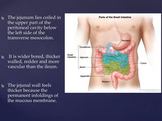
 The jejunum lies coiled in
the upper part of the
peritoneal cavity below
the left side of the
transverse mesocolon.
 It is wider bored, thicker
walled, redder and more
vascular than the ileum.
 The jejunal wall feels
thicker because the
permanent infoldings of
the mucous membrane.
Gross Anatomy of the Jejunum .
• Macroscopic anatomy :
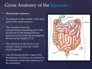
• The jejunum is the middle of the three
parts of the small intestine.
• The transition from the
extraperitoneal ascending part of the
duodenum to the intraperitoneal
jejunum occurs at the duodenojejunal
flexure (at the height of L2).
• The transition to the ileum is not
sharply marked and only visible
microscopically.
• Like all intraperitoneal organs both
the jejunum and ileum are attached to
the posterior wall of the abdomen by
the mesentery.
Jejunum
▪ Most absorption of nutrients occurs here
▪ Chemical digestion occurs here
▪ Long finger shaped villi
▪ Thick walled and wide lumen
Blood Supply
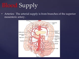
• Arteries: The arterial supply is from branches of the superior
mesenteric artery .
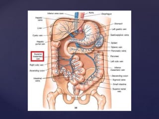
Venous Drainage of
Jejunum
• Venous drainage of jejunum is by superior mesenteric vein.
• The superior mesenteric vein reach to the point it unites
with the splenic vein and form the Portal vein(hepatic
portal vein).
• The superior mesenteric vein go with (accompanies) the
superior mesenteric artery to it’s right and anterior of the
superior mesenteric vein.
Gross anatomy of the jejunum , including blood and nerve supply, venous and lymphatic drainage.
Nerve Supply of jejunum
• Nerve Supply of jejunum derived from :
• sympathetic and parasympathetic nerves .
• The sympathetic innervation is carried by
the nerves of superior mesenteric plexus,
the parasympathetic innervation by the
vagus nerve (cranial nerve X).
Gross anatomy of the jejunum , including blood and nerve supply, venous and lymphatic drainage.
lymphatic drainage
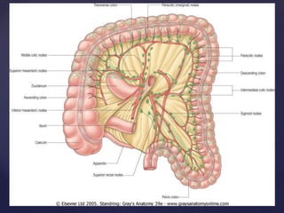
• Lymphatic vessels of the jejunum pass into mesenteric nodes via the
lacteals of the villi.
• They do not reach the superior mesenteric nodes, which the blood
vessels follow.
• All the efferents pass into the intestinal lymph trunks, which then enter
the cisterna chyli or the abdominal confluence of lymph trunks.
• The cisterna chyli drains into the thoracic duct.
mesenteric nodes
Gross anatomy of the jejunum , including blood and nerve supply, venous and lymphatic drainage.
Relations of jejunum
 The jejunum begins at
the duodenojejunal
flexure at the level of
second lumbar
vertebra.
 the duodenojejunal
flexure is located on
left of the aorta.
Location of Jejunum
 Most of the jejunum Lies in the upper part of the
abdominal cavity and to the left of the midline.
 The coils of jejunum is attached to the posterior
abdominal wall by a fan-shaped fold of
peritoneum known as the mesentery of the small
intestine.
Gross anatomy of the jejunum , including blood and nerve supply, venous and lymphatic drainage.
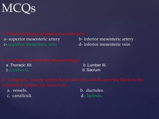
MCQs
1- Venous drainage of jejunum is drawn by :
a- superior mesenteric artery b- inferior mesenteric artery
c- superior mesenteric vein d- inferior mesenteric vein
2- At what level does the jejunum begin :
a .Thoracic XII. b .Lumber III.
. c .Lumber II. d .Sacrum.
3- Lymphatic vessels within the bowel villi, which carry the lipids to the
circulatory system, are known as:
a. vessels. b. ductules.
c. canaliculi. d. lacteals.
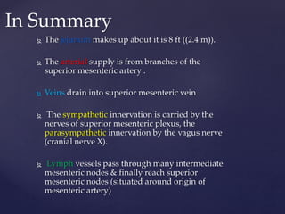
 The jejunum makes up about it is 8 ft ((2.4 m)).
 The arterial supply is from branches of the
superior mesenteric artery .
 Veins drain into superior mesenteric vein
 The sympathetic innervation is carried by the
nerves of superior mesenteric plexus, the
parasympathetic innervation by the vagus nerve
(cranial nerve X).
 Lymph vessels pass through many intermediate
mesenteric nodes & finally reach superior
mesenteric nodes (situated around origin of
mesenteric artery)
In Summary
THANK YOU

More Related Content

Gross anatomy of the jejunum , including blood and nerve supply, venous and lymphatic drainage.

  • 1. { Gross Anatomy of the Jejunum including blood and nerve supply, venous and lymphatic drainage.
  • 2. 1 ‫شديد‬ ‫رفيق‬ ‫بن‬ ‫مصطفى‬ ‫بن‬ ‫عاصم‬ 435032467 2 ‫الدايل‬ ‫عبدالرحمن‬ ‫بن‬ ‫يوسف‬ ‫بن‬ ‫عبدالرحمن‬ 435031641 3 ‫الغامدي‬ ‫لوفه‬ ‫آل‬ ‫عبدهللا‬ ‫بن‬ ‫سعود‬ ‫بن‬ ‫عبدهللا‬ 435031674 4 ‫العتيبي‬ ‫الغبيوي‬ ‫تركي‬ ‫بن‬ ‫بندر‬ ‫بن‬ ‫فراس‬ 435031634 5 ‫الشريف‬ ‫الحامد‬ ‫عبدهللا‬ ‫بن‬ ‫غالب‬ ‫بن‬ ‫فهد‬ 435032389 6 ‫المطيري‬ ‫الصيعري‬ ‫عائض‬ ‫بن‬ ‫عيد‬ ‫بن‬ ‫فيصل‬ 435031637 7 ‫الحربي‬ ‫أحمد‬ ‫بن‬ ‫موسى‬ ‫بن‬ ‫متعب‬ 435032375 8 ‫الحارثي‬ ‫الهميلي‬ ‫عبدهللا‬ ‫بن‬ ‫عيضه‬ ‫بن‬ ‫محمد‬ 435032399 9 ‫الحربي‬ ‫مشعل‬ ‫عبدالرحمن‬ 435031643 Teamwork :
  • 3.  Overview of Jejunum  Gross Anatomy of the Jejunum .  Blood Supply .  Venous Drainage .  Nerve Supply .  lymphatic drainage .  Relations of Jejunum .  Location of Jejunum .  MCQ  In Summary . Topic objectives:
  • 4. • The jejunum is the second part of the small intestine . • It is connecting the duodenum to the ileum , it is about 8 ft (2.4 m). • The jejunum begins at the duodenojejunal flexure. Overview of Jejunum
  • 5.  The jejunum lies coiled in the upper part of the peritoneal cavity below the left side of the transverse mesocolon.  It is wider bored, thicker walled, redder and more vascular than the ileum.  The jejunal wall feels thicker because the permanent infoldings of the mucous membrane.
  • 6. Gross Anatomy of the Jejunum . • Macroscopic anatomy : • The jejunum is the middle of the three parts of the small intestine. • The transition from the extraperitoneal ascending part of the duodenum to the intraperitoneal jejunum occurs at the duodenojejunal flexure (at the height of L2). • The transition to the ileum is not sharply marked and only visible microscopically. • Like all intraperitoneal organs both the jejunum and ileum are attached to the posterior wall of the abdomen by the mesentery.
  • 7. Jejunum ▪ Most absorption of nutrients occurs here ▪ Chemical digestion occurs here ▪ Long finger shaped villi ▪ Thick walled and wide lumen
  • 8. Blood Supply • Arteries: The arterial supply is from branches of the superior mesenteric artery .
  • 9. Venous Drainage of Jejunum • Venous drainage of jejunum is by superior mesenteric vein. • The superior mesenteric vein reach to the point it unites with the splenic vein and form the Portal vein(hepatic portal vein). • The superior mesenteric vein go with (accompanies) the superior mesenteric artery to it’s right and anterior of the superior mesenteric vein.
  • 11. Nerve Supply of jejunum • Nerve Supply of jejunum derived from : • sympathetic and parasympathetic nerves . • The sympathetic innervation is carried by the nerves of superior mesenteric plexus, the parasympathetic innervation by the vagus nerve (cranial nerve X).
  • 13. lymphatic drainage • Lymphatic vessels of the jejunum pass into mesenteric nodes via the lacteals of the villi. • They do not reach the superior mesenteric nodes, which the blood vessels follow. • All the efferents pass into the intestinal lymph trunks, which then enter the cisterna chyli or the abdominal confluence of lymph trunks. • The cisterna chyli drains into the thoracic duct.
  • 16. Relations of jejunum  The jejunum begins at the duodenojejunal flexure at the level of second lumbar vertebra.  the duodenojejunal flexure is located on left of the aorta.
  • 17. Location of Jejunum  Most of the jejunum Lies in the upper part of the abdominal cavity and to the left of the midline.  The coils of jejunum is attached to the posterior abdominal wall by a fan-shaped fold of peritoneum known as the mesentery of the small intestine.
  • 19. MCQs 1- Venous drainage of jejunum is drawn by : a- superior mesenteric artery b- inferior mesenteric artery c- superior mesenteric vein d- inferior mesenteric vein 2- At what level does the jejunum begin : a .Thoracic XII. b .Lumber III. . c .Lumber II. d .Sacrum. 3- Lymphatic vessels within the bowel villi, which carry the lipids to the circulatory system, are known as: a. vessels. b. ductules. c. canaliculi. d. lacteals.
  • 20.  The jejunum makes up about it is 8 ft ((2.4 m)).  The arterial supply is from branches of the superior mesenteric artery .  Veins drain into superior mesenteric vein  The sympathetic innervation is carried by the nerves of superior mesenteric plexus, the parasympathetic innervation by the vagus nerve (cranial nerve X).  Lymph vessels pass through many intermediate mesenteric nodes & finally reach superior mesenteric nodes (situated around origin of mesenteric artery) In Summary