15 dural venous sinuses
•Download as PPTX, PDF•
67 likes•18,047 views

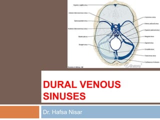
1) The document describes the dural venous sinuses, which drain blood from the brain into the internal jugular veins. They include the superior and inferior sagittal sinuses, straight sinus, transverse sinus, sigmoid sinus, occipital sinus, cavernous sinus, and superior and inferior petrosal sinuses. 2) The dural sinuses have no valves or muscle in their walls and are located between the layers of the dura mater. They drain blood from the brain and cerebrospinal fluid from the subarachnoid space. 3) The blood in the dural sinuses ultimately drains into the internal jugular veins in the neck through a series of sinuses and


15 dural venous sinuses
- 2. DURAL VENOUS SINUSES Dr. Hafsa Nisar
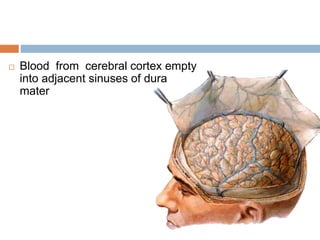
- 3. Blood from cerebral cortex empty into adjacent sinuses of dura mater
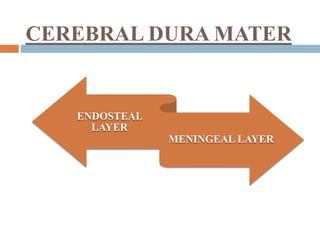
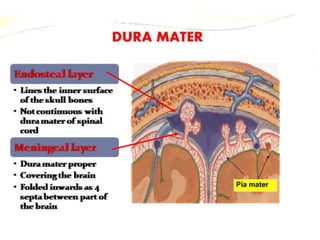
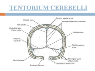
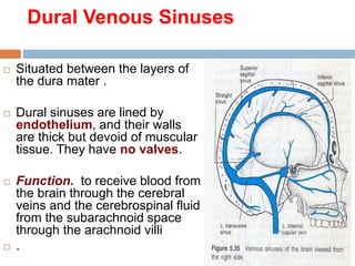
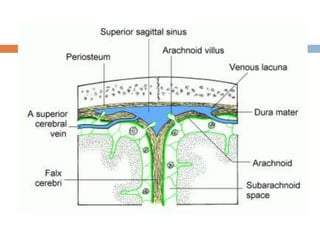
- 7. Dural Venous Sinuses Situated between the layers of the dura mater . Dural sinuses are lined by endothelium, and their walls are thick but devoid of muscular tissue. They have no valves. Function. to receive blood from the brain through the cerebral veins and the cerebrospinal fluid from the subarachnoid space through the arachnoid villi .
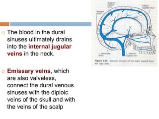
- 9. The blood in the dural sinuses ultimately drains into the internal jugular veins in the neck. Emissary veins, which are also valveless, connect the dural venous sinuses with the diploic veins of the skull and with the veins of the scalp
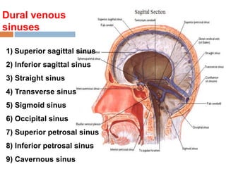
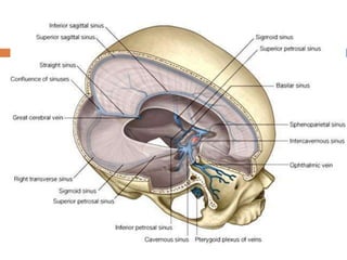
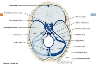
- 10. Dural venous sinuses 1) Superior sagittal sinus 2) Inferior sagittal sinus 3) Straight sinus 4) Transverse sinus 5) Sigmoid sinus 6) Occipital sinus 7) Superior petrosal sinus 8) Inferior petrosal sinus 9) Cavernous sinus
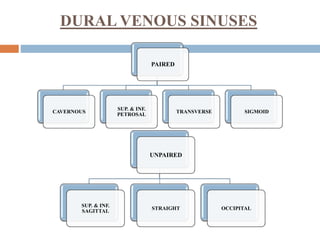
- 11. DURAL VENOUS SINUSES PAIRED CAVERNOUS SUP. & INF. PETROSAL TRANSVERSE SIGMOID UNPAIRED SUP. & INF. SAGITTAL STRAIGHT OCCIPITAL
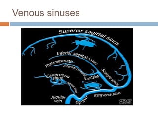
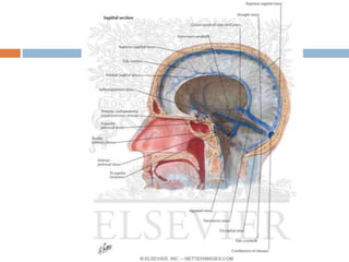
- 12. Venous sinuses
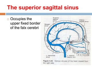
- 13. The superior sagittal sinus Occupies the upper fixed border of the falx cerebri
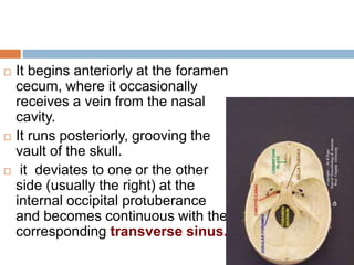
- 14. It begins anteriorly at the foramen cecum, where it occasionally receives a vein from the nasal cavity. It runs posteriorly, grooving the vault of the skull. it deviates to one or the other side (usually the right) at the internal occipital protuberance and becomes continuous with the corresponding transverse sinus.
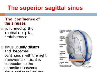
- 15. The superior sagittal sinus The confluence of the sinuses is formed at the internal occipital protuberance. sinus usually dilates and becomes continuous with the right transverse sinus; it is connected to the opposite transverse
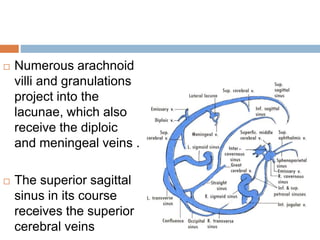
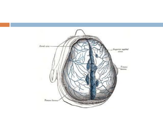
- 16. Numerous arachnoid villi and granulations project into the lacunae, which also receive the diploic and meningeal veins . The superior sagittal sinus in its course receives the superior cerebral veins
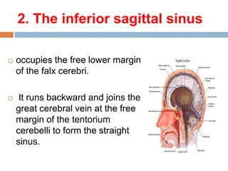
- 18. 2. The inferior sagittal sinus occupies the free lower margin of the falx cerebri. It runs backward and joins the great cerebral vein at the free margin of the tentorium cerebelli to form the straight sinus.
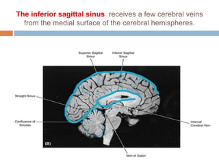
- 20. The inferior sagittal sinus receives a few cerebral veins from the medial surface of the cerebral hemispheres.
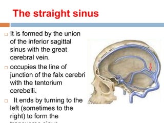
- 21. The straight sinus It is formed by the union of the inferior sagittal sinus with the great cerebral vein. occupies the line of junction of the falx cerebri with the tentorium cerebelli. It ends by turning to the left (sometimes to the right) to form the
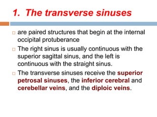
- 22. 1. The transverse sinuses are paired structures that begin at the internal occipital protuberance The right sinus is usually continuous with the superior sagittal sinus, and the left is continuous with the straight sinus. The transverse sinuses receive the superior petrosal sinuses, the inferior cerebral and cerebellar veins, and the diploic veins.
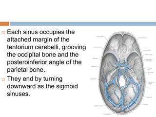
- 23. Each sinus occupies the attached margin of the tentorium cerebelli, grooving the occipital bone and the posteroinferior angle of the parietal bone. They end by turning downward as the sigmoid sinuses.
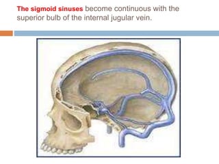
- 24. The sigmoid sinuses are a direct continuation of the transverse sinuses. Each sinus turns downward and medially and grooves the mastoid part of the temporal bone. pass inferiorly through the posterior part of the jugular foramen to become continuous with the superior bulb of the internal jugular vein.
- 27. The sigmoid sinuses become continuous with the superior bulb of the internal jugular vein.
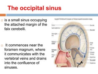
- 28. The occipital sinus is a small sinus occupying the attached margin of the falx cerebelli. It commences near the foramen magnum, where it communicates with the vertebral veins and drains into the confluence of sinuses.
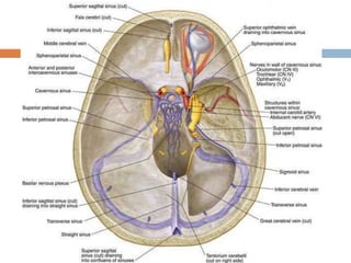
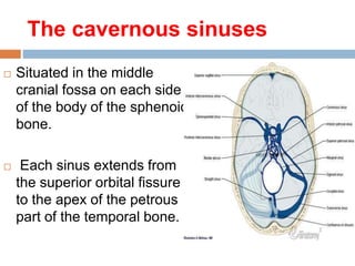
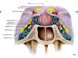
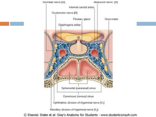
- 29. The cavernous sinuses Situated in the middle cranial fossa on each side of the body of the sphenoid bone. Each sinus extends from the superior orbital fissure to the apex of the petrous part of the temporal bone.
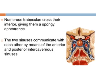
- 30. Numerous trabeculae cross their interior, giving them a spongy appearance. The two sinuses communicate with each other by means of the anterior and posterior intercavernous sinuses,
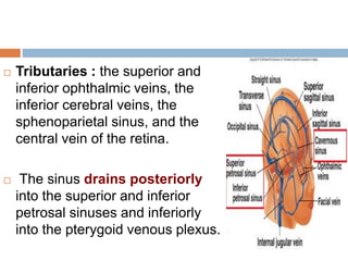
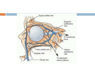
- 31. Tributaries : the superior and inferior ophthalmic veins, the inferior cerebral veins, the sphenoparietal sinus, and the central vein of the retina. The sinus drains posteriorly into the superior and inferior petrosal sinuses and inferiorly into the pterygoid venous plexus.
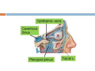
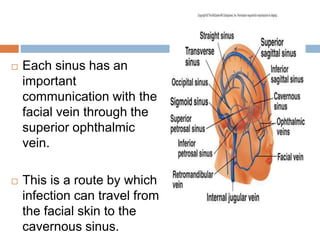
- 34. Each sinus has an important communication with the facial vein through the superior ophthalmic vein. This is a route by which infection can travel from the facial skin to the cavernous sinus.
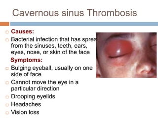
- 39. Cavernous sinus Thrombosis Causes: Bacterial infection that has spread from the sinuses, teeth, ears, eyes, nose, or skin of the face Symptoms: Bulging eyeball, usually on one side of face Cannot move the eye in a particular direction Drooping eyelids Headaches Vision loss
- 40. Tests include: CT scan of the head Magnetic resonance imaging (MRI) of the brain Magnetic resonance venogram Sinus x-ray Treatment: Cavernous sinus thrombosis is treated with high-dose antibiotics given through a vein (IV). Sometimes surgery is needed to drain
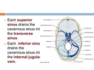
- 41. Petrosal sinuses The superior and inferior petrosal sinuses are small sinuses situated on the superior and inferior borders of the petrous part of the temporal bone on each side of the skull.
- 42. Each superior sinus drains the cavernous sinus into the transverse sinus Each inferior sinus drains the cavernous sinus into the internal jugular vein.
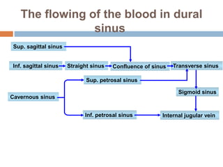
- 43. The flowing of the blood in dural sinus Sup. sagittal sinus Inf. sagittal sinus Straight sinus Confluence of sinus Transverse sinus Cavernous sinus Sup. petrosal sinus Inf. petrosal sinus Internal jugular vein Sigmoid sinus