Showing posts with label CT scan. Show all posts

Saturday, November 23, 2024

Chest (or abdominal?) pain and ECG artifact.

An elderly woman presented with one day of chest and right arm pain, and also abdominal pain.  There was associated tingling and numbness in the right hand and generalized weakness, worse on the right side.

A triage ECG was recorded:


Smith: there is widespread artifact, except in lead III.  Since lead III is not artifactual, one can deduce that the artifact is caused by movement of the right arm electrode, so that electrode should be moved and the ECG re-recorded.  This makes it difficult to assess. 

But one can see that there is some ST depression in lead III, which should always make you think that it is reciprocal to some, often minimal, STE in aVL (of high lateral OMI).  Thus, aVL, I, and V2 must be scrutinized, but they are distorted by artifact.  

This is extremely easy to overlook, unless there is a high suspicion of OMI.

Single limb lead electrode artifact is explained here, in a case of Arterial Pulse Tapping Artifact (APTA): Bizarre (Hyperacute??) T-waves


Clinical course: The provider recognized artifact, but did not see anything worrisome and regrets not ordering another.

Labs, including troponins, were ordered and the patient needed be placed in the waiting room pending a bed.

When the provider next looked up lab results, it was noticed that the first troponin was 45,000 ng/L.  This is then a large MI, but it is subacute.

There was a previous ECG available for review:
This shows baseline STE in inferior leads, including lead III.
At the time is was recorded, there was no a myocardial infarction; this was baseline STE (normal variant)
This previous ECG confirms that the STD in lead III is very abnormal


Due to the very high troponin, the patient was brought to the critical care area and another ECG was recorded:
Sinus rhythm with PVCs
There is inferior STD with STE and a coved ST segment in aVL.  There is STE in V5-6.  There are new Q-waves in aVL, V5-6.  
It is diagnostic of OMI, but this is SUBACUTE OMI

I sent this ECG to my "EKG Nerdz" friends, without any clinical info at all and they answered "OMI"


The Queen said: "STEMI-Equivalent with High Confidence:"

Notice she sees findings in both normal beats and PVCs.


There was some question of whether the patient was having abdominal pathology, and she also had a history of aortic pathology, so a chest abd/pelvic with aorta angiogram was ordered.

If this were ACUTE (vs. SUBACUTE) OMI, that would result in an undesirable delay.

But this is clearly a subacute MI, with most of the damage done.  How do I know?

1) Very high initial troponin of 45,000 ng/L

2) A full day of chest pain

3) Q-waves on the ECG, with some T-wave inversion

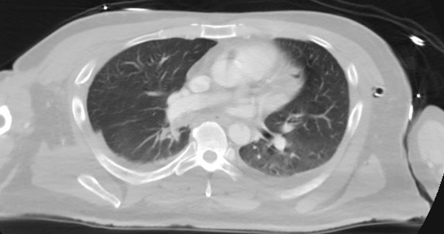
Here is one frame of the CT scan which includes the heart:

Can you spot the infarct?


Here is an arrow pointing to it:


The Cath Lab was activated.

Angiogram:

100% occluded ramus intermedius.  The ramus is an occasional artery between the circumflex and the LAD, and often takes the place of a large first diagonal, and has the same distribution.

It was opened and stented.

Here are the troponins:


Echo:

Normal LV size and hyperdynamic systolic function with an estimated EF of 77%.

Regional wall motion abnormality--mid anterior akinesis.


Compared to TTE from 7/3/24:  the anterior regional wall motion abnormality is new and is consistent with ischemia/infarction in the LAD territory


===================================

MY Comment, by KEN GRAUER, MD (11/23/2024):

===================================
There are several insightful aspects of today's case.
  • Marked artifact in the initial ECG (TOP tracing in Figure-1) — makes assessment of the limb leads difficult — since lead III (within the BLUE rectangleis the only undistorted lead. But in a patient with CP (Chest Pain) — the T wave in this single undistorted limb lead clearly looks to be hyperacute (disproportionately "bulky" considering modest size of the QRS in this lead)
  • I next looked at leads V2,V3,V4 for sign of posterior OMI — since CP + hyperacute T wave in lead III + suggestion of posterior OMI would be enough to greatly enhance my confidence of acute OMI in this initial tracing. Unfortunately — artifact distortion of the the ST segments in these 3 chest leads prevents drawing any conclusions. But, in this patient with CP — ECG #1 needs to be immediately repeated!
  • As per Dr. Smith — the fact that artifact in ECG #1 is maximal in leads I and II (with lead III undistorted) — points to the RA extremity as the "culprit" (See My Comment in the December 5, 2022 post of Dr. Smith's ECG Blog for review on how to determine the "culprit" extremity within seconds).

Figure-1: Comparison between the initial ECG and the repeat ECG in today's case.

The 2nd ECG:
Assessment of the next ECG in today's case (BOTTOM tracing in Figure-1) — provides its own set of insightful observations:
  • Overall — the artifact is decidedly less in ECG #2. That said — Note how the "culprit" extremity has changed! Here limb lead artifact is maximal in leads IIII and aVL — with lead II no more than minimally affectedNote also that instead of the artifact distorting the entire recording of 5/6 limb leads (as it did in ECG #1) — the artifact distortion in ECG #2 is primarily of the baseline! This localizes the source of artifact to the LA electrode (rather than pointing to a tremulous RA extremity — as was the case in ECG #1, in which large amplitude artifactual deflections were seen throughout in the affected limb lead recordings).

Although the artifact in ECG #2 should also prompt repeating the ECG — we nevertheless can draw important conclusions from this 2nd tracing.
  • The rhythm is ventricular bigeminy (each of the even-numbered beats in ECG #2 is a PVC).
  • That every-other-beat in ECG #2 is a PVC (and not a PAC conducted with RBBB aberration) — can be established by the fact that underlying sinus P waves continue throughout the tracing (YELLOW arrows in the long lead II rhythm strip showing on-time sinus P waves producing subtle distortion of the beginning of the ST segment of each PVC). In addition, the direction of the initial deflection in 4 of the chest leads is different for sinus beats and the PVCs (whereas sinus beats #9,11,13 in leads V1-thru-V4 all manifest an initial R wave — beas #8,10,12 all manifest an initial negative deflection).

PEARL:
 As we have occasionally seen in other cases in Dr. Smith's ECG Blog — it is the PVCs (and not the sinus-conducted beats) that provide more incriminating evidence of an ongoing acute event.
  • Consider lead aVL (within the PURPLE rectangle) — in which the most markedly abnormal ST elevation is seen in beats #4 and #6 (with the tricky aspect of beat #4 being the lead change marker that hides the QRS of beat #4).
  • Lead I appears to show abnormal ST elevation in both sinus beats and the PVC in this lead — although artifact make assessment difficult in this lead.
  • But BLUE arrows in lateral chest leads V5 and V6 show what appears to be disproportionately elevated J-point ST elevation in the PVC ( = beat #12).

================================  
Links to Examples of ARTIFACT:
Artifact is a reality of clinical practice — so comfort in assessing tracings with artifact is essential. What follows below is my expanding list of technical "misadventures" — most from Dr. Smith's ECG Blog — some from other sources (NOTE: As I did not previously keep track of these — there are additional examples of artifact sprinkled through Dr. Smith's ECG Blog that I have not yet included here ... ).


Sunday, April 9, 2023

Off and on chest pain for 24 hours in a 50s year old man

Submitted by Ali Khan MD and James Mantas MD, MS, written by Pendell Meyers


A man in his 50s with history of diabetes, hypertension, and tobacco use presented to the ED with 24 hours of worsening left sided chest pain radiating to the back, characterized as squeezing and pinching, associated with shortness of breath. His pain was initially mild, then became severely worse several hours prior to presentation, but then eased off again and was minimal on arrival. There was no associated diaphoresis, nausea, vomiting, arm pain, jaw pain, syncope, lightheadedness or other acute symptoms.

Initial vitals: Temp 36.7 C, BP 161/79, RR 16, HR 70, Pulse Ox 97%


Initial ECG (during "minimal" pain):

What do you think?


Meyers: I was sent this ECG with zero clinical information (and without the prior ECG below), and at that time I said that I did not see evidence of OMI. Yet there is minimal STE in III, some STD in I and aVL, and terminal T wave inversion in III, suggesting some reperfusion and consistent with the resolution of most of the pain. I should have seen that this could be inferior OMI with current reperfusion. If I had the history or the prior ECG below, I may have arrived at that diagnosis.

PM Cardio Queen of Hearts AI said "Not OMI - High Confidence". She is not able to compare to prior ECGs (yet).


Prior ECG on file:

Normal ECG, showing that the features above are new.


He was treated initially with 325mg of ASA and 0.4mg SL NTG.


Bedside POCUS revealed inferior wall motion abnormality (images not available).

Smith comment: this is clearly an inferior OMI with reperfusion.  Does it help to know there is a wall motion abnormality?  Only if you are uncertain about the diagnosis of ACS.   Reperfusion will usually NOT restore cardiac wall motion.  The myocardium is "stunned" and will often (not always!!) remain hypo- or akinetic for some time even if not infarcted, just due to the profound ischemia incurred during the occlusive phase.  Even many NOMI have wall motion abnormalities.  Thus, an acute wall motion abnormality is not a sign of active or persistent ischemia, and thus is not necessarily an indication for emergent cath. 

The EM physicians activated their "heart alert" pathway, which immediately summons the cardiology fellow to bedside to evaluate for potential emergent cardiac catheterization lab activation.

The interventional cardiologist was not initially convinced this was ACS and recommended CT pulmonary angiography to rule out pulmonary embolism.

The pain returned/worsened, and the patient was then started on a nitroglycerin drip. Given the patients persistent and worsening pain and radiation to the back the team opted for a CT Aortogram.


Here is the ECG with returning/worsening pain:

Increasing STE in III and aVF, increasing reciprocal STD in I and aVL.
Inferior OMI until proven otherwise.
OMI AI says "OMI - Low confidence" on this ECG.


High sensitivity troponin T, in ng/L: 
127 (12:00pm), 154 (1:05pm), 206 (2:25pm), none further measured.

Smith comment: an initial hs troponin (I or T) above 50 ng/L in a chest pain patient without a history of elevated troponin, cardiomyopathy, or end-stage renal disease has a very high positive predictive value for Acute MI (which in this case would be a type I MI), but does not differentiate between OMI and Non-OMI.  (Large PE is also possible but this does NOT sound like a PE case).  A delta is hardly necessary for the diagnosis and there is no delta which can differentiate OMI from NOMI.  Therefore, a patient with persistent pain and a troponin at this level has Acute MI with ongoing ischemia and should go to the cath lab regardless of the ECG (this is by American and European guidelines).

After the 2nd troponin revealed a significant delta and given patients persistent pain a Heparin drip was started.


CT Aortogram revealed: No pulmonary embolism, No aortic dissection. "Decreased perfusion within the posterior descending artery territory suspicious for developing infarct."

Here is a screenshot of the image in question:

The arrow points to the area of ischemia.  Subtle!


Angiogram happened soon after:

LAD (mid): 80% stenosis

OM1 (prox): 80%

Ramus (ostial): 80% stenosis

RCA: 99% stenosis, no TIMI flow reported, felt to be culprit lesion, but for some technical reason unable to perform PCI and deemed more favorable for delayed CABG.


I am not sure whether the patient had pain at the time of cath, but I suspect so.

Overnight the chest pain persisted, and ECG showed continued progression of OMI post cath (no intervention done):


Formal echo:

Hypokinesis of the inferior wall

EF 69% with globally normal systolic function

Normal LV thickness

No pericardial effusion


Clinical course:

1 day after admission: Intra-aortic balloon pump placed, exact indication uncertain

2 days after admission: 3 vessel CABG performed by Cardiothoracic Surgery: Venous grafting of OM and Ramus, LIMA to LAD, unable to intervene on RCA.

The patient survived to discharge, but long-term follow up is unavailable.


No peak troponin was measured, and no TIMI flow was recorded, so it is not 100% definite that this case would meet the OMI definitions we have used in our studies so far. The culprit lesion with the ECGs above make me almost certain that this case meets the conceptual definition of OMI.


==================================
My Comment by KEN GRAUER, MD (4/9/2023):
==================================
The KEY point I'd like to emphasize regarding today's case is that specific diagnosis of the "culprit" artery with acute OMI is less important than simple recognition that an acute cardiac event is ongoing.

  • To illustrate this point — I've put the first 2 ECGs in today's case together in Figure-1.


My Initial Thoughts on Today's Case:
Like Dr. Meyers — I first saw ECG #1 without the benefit of any clinical history, other than knowing that this tracing was recorded in the ED. 
  • Most of the tracings we review from the ED are recorded in patients with some sort of chest pain history — though we were sent this tracing without knowing whether this CP (Chest Pain) was acute and ongoing — vs — present for several days and now resolved — vs — non-cardiac-sounding CP — vs — a "routine" ECG done for some other emergency disorder (ie, an acute abdomen) in a patient about to be taken to the OR.

  • My Initial Reply regarding ECG #1: Is this ECG from a patient with CP? If so — I'm suspicious of recent infero-postero OMI. That said — I agreed completely with Dr. Meyers that ECG #1 was not specific for OMI.
  • That said — as soon as I learned this patient's CP had significantly decreased (and was minimal) at the time ECG #1 was recorded — I felt my initial impression was supported, and that ECG #1 certainly could be consistent with recent OMI, now with reperfusion ST-T waves. Additional information would be needed for confirmation (ie, serial troponins — follow-up tracings, as well as search for a prior ECG— but the onus was clearly on emergency providers to prove this tracing was not reflective of an acute ongoing event, rather than the other way around.

Figure-1: Comparison of the first 2 ECGs in today's case. (To improve visualization — I've digitized the original ECG using PMcardio).


What Does the Initial ECG Show?
The initial ECG in today's case clearly suggests that at some point in time — the patient has had an inferior infarction:
  • The Q wave in lead III of ECG #1 is huge. There is also a significant Q wave in lead aVF — that is equal in depth to the height of the tiny R wave in this lead. 
  • As per Dr. Meyers — there is slight ST elevation in lead III, with terminal T wave inversion — as well as potential reciprocal changes (ST flattening and slight depression) in high-lateral leads I and aVL.

Subtle ST-T wave abnormalities are seen in virtually all of the remaining leads:
  • There is nonspecific straightening of the ST segment in lead II.
  • There is early transition — with abrupt appearance of a predominant R wave already by lead V2 (consistent with posterior MI at some point in time).
  • Instead of gradually upsloping ST segments with upright T waves in leads V2-thru-V6 — T waves are barely seen, with nonspecific ST segment flattening in these leads (and with slight ST depression in lead V4).

BOTTOM Line for ECG #1:
  • Prior inferior (and probably also posterior) infarction has occurred at some point in time. 
  • ECG #1 is not diagnostic of acute occlusion. That said, given the history in this 50ish year old man with risk factors, who describes 24 hours of worsening CP — with near resolution of CP at the time he finally arrived in the ED — ECG #1 is clearly consistent with a possible new infero-postero OMI, now with spontaneous reperfusion corresponding to the time the patient reports the decrease in CP (ie, In between the period of acute ST elevation and reperfusion T wave inversion — can be a phase of "pseudo-normalization", during which ST-T waves manifest diffuse flattening — but not much ST elevation or depression).

  • Alternatively — ECG #1 might represent multi-vessel disease, on top of which there may (or may not be) ECG changes of another new event.


Benefit of the Prior Tracing:
Lead-by-lead comparison of ECG #1 with the prior ECG on file proved insightful:
  • Q waves were previously present in leads III and aVF — albeit compared to R wave amplitude in these leads in the prior tracing — the Q waves in leads III and aVF of ECG #1 are now larger.
  • ST-T wave appearance in each of the inferior leads is clearly different in these 2 tracings. A normal ST-T wave was previously seen in lead II. There was no ST elevation in lead III of the prior tracing, and the T wave was all negative instead of biphasic (up-down).
  • ST-T waves looked normal in leads I and aVL of the prior tracing.
  • In the chest leads — transition was not as abrupt (ie, a predominant R wave was not seen until lead V3 in ECG #2). There was no ST depression in lead V4 — and precordial lead T waves looked uniformly better in the prior tracing.

  • IMPRESSION after the Prior Tracing was Found: There can no longer be any doubt that ST-T wave changes in ECG #1 are acute. Prompt cath was indicated at this point in the case.


Final POINTS:
  • Even "after the fact" — I have trouble identifying the specific "culprit" artery from review of the initial ECG and the prior tracing on file — and, difficulty distinguishing the picture painted by these 2 ECGs from what might be seen with acute ischemia and multi-vessel disease. Clinically, this does not matter — since regardless, the patient's 24-hour history of CP in the context of obvious new ECG changes should merit prompt cath as soon as the prior ECG was found.

  • P.S. — One wonders what took this patient with longstanding diabetes, hypertension and tobacco use a full 24 hours to make it to the ED. This patient is lucky he made it to the ED alive.


Friday, November 25, 2022

Patient in Single Vehicle Crash: What is this ST Elevation, with Peak Troponin of 6500 ng/L?

A 30-something male was involved in a single vehicle crash and had multiple injuries. 

As a routine part of his critical trauma evaluation, he had an ECG recorded:

There is an rSR" in V1 and V2, with downsloping ST segment and inverted T-wave which is very similar to a Brugada Type 1 phenocopy.   I was shown this ECG and thought that it could perhaps be Brugada, but I was more suspicious for Right Ventricular (RV) myocardial contusion.  The RV is the most anterior part of the chest and is most likely to be contused with anterior chest trauma.  Moreover, this degree of ST Elevation is very unusual for Brugada.


On the other hand, the accident was unexplained.  Did the patient have an episode of VT due to Brugada, and lose consciousness while driving?


Here is one slice of the patient's chest CT

Note the pneumothorax, but the pneumothorax underlies leads V3-V5, not V1 and V2.  

On the other hand, the RV lies directly under leads V1 and V2; this makes pneumothorax less likely as an etiology of the abnormal ECG.


The pneumothorax was expanded with a chest tube


At 17 hours, another ECG was recorded:

It is now much less dramatic and has the morphology of Type 2 Brugada


The hs troponin I peaked at 6500 ng/L -- this strongly suggests myocardial contusion.  Is there also Brugada?


An echocardiogram was done.  Here is the result:

The estimated left ventricular ejection fraction is 50 %.

There is no left ventricular wall motion abnormality identified.

Right ventricular prominence.

Normal right ventricular systolic performance lower limits of normal.


At 40 hours, another ECG was recorded:

Again, it has features of Type 1 Brugada


The electrophysiologist who was consulted was concerned for Brugada:

"It is unclear what precipitated his motor vehicle collision.  The patient is not able to recall events preceding the crash (syncope, dizziness, lightheadedness).  He has a family history concerning for arrhythmia.  Given the circumstances of his car crash, we presume it was due to an underlying arrhythmia.  I suspect his troponin elevation is due to acute myocardial injury (not acute thrombus) related to his polytrauma, possibly cardiac contusion, and reassuring that he has no wall motion abnormalities on TTE."


An MRI was done:  


1) Normal LV function with no wall motion abnormalities
2) Normal dimensions of all cardiac chambers
3) No evidence of left ventricular myocardial scar on delayed enhancement
sequences after contrast administration
4) Hypokinesis involving the free wall of the right ventricle with delayed enhancement, concerning for right ventricular contusion. This appears to be immediately beneath the sternal fracture.   At this point, recommend a short-term follow-up cardiac MRI tailored to evaluation of the right ventricle, in a few days after patient recovery.


2 weeks


Here is the final electrophysiology note:


It is unclear what precipitated his motor vehicle collision.  The patient is not able to recall events preceding the crash (syncope, dizziness, lightheadedness, though reports feeling a popping sound).  He has a family history concerning for arrhythmia with his father requiring some sort of device (PPM, ICD, unclear) at a young age. He has Brugada pattern on EKG with possible syncope, which is concerning for underlying paroxysmal ventricular arrhythmia.  However, his cardiac MRI shows scar that is also possibly associated with his sternal fracture given his location.  


Given the possibility of a ventricular arrhythmia, we would recommend the patient receive a Zoll LifeVest prior to discharge and repeat the cardiac MRI in 2-3 weeks.


Pending the results of the MRI, he could be considered for ICD placement if it does seem more consistent with Brugada syndrome rather than injury to the RV from contusion.  In the meantime will use LifeVest to bridge him during ongoing workup for his RV scar.


Diagnosis: 

1. Definite Right Ventricular Myocardial Contusion and 

2. Possible Underlying Brugada syndrome.


If and when I get a confirmation or refutation of Brugada syndrome, I will update this post.


See our other cases of myocardial contusion and related cases (some of which have an important diagnosis OTHER THAN myocardial contusion!):


===================================

MY Comment, by KEN GRAUER, MD (11/25/2022):

===================================

Highly interesting post by Dr. Smith regarding a 30-something male with multiple injuries from a motor vehicle accident. The KEY issues are highlighted by Dr. Smith's title of this post = "What is this ST Elevation, with Peak Troponin of 6500 ng/L?"

When I first saw the initial ECG in today's case — I interpreted the ECG changes in leads V1,V2 as consistent with a Brugada-1 pattern, most likely Brugada Phenocopy
  • As we've discussed on numerous other posts in Dr. Smith's ECG Blog (See My Comment at the bottom of the page in the May 5, 2022 post) —  a growing number of conditions other than Brugada Syndrome have been found to temporarily produce a Brugada-1 ECG pattern. These include (among others) — acute febrile illness — variations in autonomic tone — hypothermia — ischemia-infarction — malignant arrhythmias — cardiac arrest — and especially Hyperkalemia. Patients with such conditions that may transiently mimic the ECG findings of a Brugada-1 pattern are said to have Brugada Phenocopy. The importance of being aware of this phenomenon of Brugada Phenocopy — is that correction of the underlying condition may result in resolution of the Brugada-1 ECG pattern, with a much better prognosis compared to patients with true Brugada Syndrome.

  • Kazmi et al have reported on a case in which chest trauma was transiently associated with development of a Brugada-1 ECG pattern (J Am Coll Cardiol 73 [9-Supp-1], 2019). The authors postulate that the mechanism for producing the Brugada-1 ECG pattern was that anterior chest wall trauma caused a non-homogeneous depolarizing current (possibly related to opening of mechanicallly sensitive cardiac K+ATP channels). This ECG abnormality was transient in the case they report — consistent with trauma-induced Brugada Phenocopy.

  • Alternatively (as per Dr. Smith) — it could be that a malignant arrhythmia that altered consciousness was the precipitating cause of both the motor vehicle crash, and of the Brugada-1 pattern on ECG.


As noted above — Dr. Smith was especially suspicious of RV (Right Ventricular) Myocardial Contusion as primarily responsible for the patient's initial ECG pattern.
  • While impossible to rule out a malignant arrhythmia as the cause of the motor vehicle accident — I think it insightful to consider the combination of Brugada-1 Phenocopy + RV Myocardial Contusion as both contributing to the serial ECG changes seen in today's case.

What are the ECG Findings of Cardiac Contusion?
I've copied KEY points from My Comment in the August 6, 2022 post in Dr. Smith's ECG Blog —  regarding the answer to this question. Overall — the ECG is less than optimally sensitive for detecting cardiac injury following blunt trauma. This is because the anterior anatomic position of the RV (Right Ventricle), and its immediate proximity to the sternum — makes the RV much more susceptible to blunt trauma injury than the LV. But because of the much greater electrical mass of the LV — electrical activity (and therefore ECG abnormalities) from the much smaller and thinner RV are more difficult to detect. To REVIEW (Sybrandy et al: Heart 89:485-489, 2003 — Alborzi et al: J The Univ Heart Ctr 11:49-54, 2016 — and Valle-Alonso et al: Rev Med Hosp Gen Méx 81:41-46, 2018) — ECG findings commonly reported in association with Cardiac Contusion include the following:
  • None (ie, The ECG may be normal — such that not seeing any ECG abnormalities does not rule out the possibility of cardiac contusion).
  • Sinus Tachycardia (common in any trauma patient ...).
  • Other Arrhythmias (PACs, PVCs, AFib, Bradycardia and AV conduction disorders — potentially lethal VT/VFib).
  • RBBB (as by far the most common conduction defect — owing to the more vulnerable anatomic location of the RV). Fascicular blocks and LBBB are less commonly seen.
  • Signs of Myocardial Injury (ie, Q waves, ST elevation and/or depression — with these findings suggesting LV involvement).
  • QTc prolongation.
  • Brugada Phenocopy (as per the Kazmi et al reference that I cited above).

  • NOTE: Prediction of cardiac contusion "severity" on the basis of cardiac arrhythmias and ECG findings — is an imperfect science.

Additional KEY Points regarding Cardiac Contusion:
Despite the predominance for RV (rather than LV) injury — use of a right-sided V4R lead has not been shown to be helpful compared to use of a standard 12-lead ECG for detecting ECG abnormalities.
  • In addition to ECG abnormalities related to the blunt trauma of cardiac contusion itself — Keep in mind the possibility of other forms of cardiac injury in these patients (ie, valvular injury, aortic dissection, septal rupture) — as well as the possibility of a primary cardiac event (ie, acute MI may have been the cause of an accident that led up to the trauma).
  • ECG abnormalities may be delayed — so repeating the ECG if the 1st tracing is normal is appropriate when concerned about severe traumatic injury.
  • That said — IF troponin is normal at 4-6 hours and IF the ECG is normal — then the risk of cardiac complications is extremely low.


What About Today's Case?
As I reconsidered today's case — I think the 3 serial ECGs are consistent with both ST elevation in leads V1,V2 from acute RV myocardial contusion + variations of Brugada-1 and Brugada-2 ECG Phenocopy patterns.
  • For clarity — I've put together in Figure-1 a visual review of the 2 Brugada ECG patterns (from My Comment in the May 5, 2022 post) — with leads V1,V2,V3 from the 3 ECGs done in today's case. 

  • In ECG #1 — I still find it hard to distinguish between a Brugada-1 ECG pattern vs ST elevation in leads V1,V2 from acute RV insult vs some combination of the two. I suspect we are seeing a combined ECG effect (as well as a tall, slender R' in lead V1 from myocardial contusion-induced incomplete RBBB).

  • In ECG #2 — the ST-T wave abnormality is greatly reduced, and now primarily found in lead V2. Although bearing similarity to the Brugada-2 ECG pattern (in Panel B of Figure-1) — the shape of the ST segment descent from the r' peak is unusual for a Brugada-2 pattern in that it includes a short coved segment (outlined in RED — highlighted by the BLUE arrow). Doesn't this shape look to be more consistent with superimposed ST elevation rather than a Brugada-2 pattern alone?

  • In ECG #3 — this unusually shaped coved segment (outlined in RED — highlighted by the BLUE arrow) is now even more prominent. I thought this coved segment to be more consistent with superimposed ST elevation from acute RV injury rather than solely the result of a Brugada-1 and/or Brugada-2 pattern.

  • BOTTOM LINE: As per Dr. Smith's discussion above — the markedly elevated serum troponin — as well as additional imaging studies confirmed RV Myocardial Contusion. While I believe the serial ECGs in today's case show variations of Brugada-1 and -2 Phenocopy — I suspect the BLUE arrows in Figure-1 highlight superimposed ST elevation from acute RV injury. That said, the "good news" — is that this patient should recover (and his ECG will most probably normalize as he does!).
 
Figure-1: Visual summary of ECG Patterns in Brugada Syndrome (adapted from the article by Brugada et al in JACC: Vol. 72; Issue 9; 2018) — A) Brugada-1 ECG pattern, showing coved ST-segment elevation ≥2 mm in ≥1 right precordial lead, followed by a negative T-wave.  B) Brugada-2 ECG pattern (the “Saddle-back” pattern) — showing concave-up ST-segment elevation ≥0.5 mm (generally ≥2 mm) in ≥1 right precordial lead, followed by a positive T-wave.  C) Additional criteria for diagnosis of a Brugada-2 ECG pattern (TOPthe ß-angleBOTTOMA Brugada-2 pattern is present if 5 mm down from the maximum r’ rise point — the base of the triangle formed is ≥4).

BELOW this summary of Brugada ECG Patterns — I've put together the V1,V2,V3 leads from the 3 serial ECGs obtained in today's case (See text).


Recommended Resources