Please enter url.
Login
Logout
Please enter url.
Pleura Pulmonar Pulm N Tuberculosis Cavidad Pleural Rgano | Sexiz Pix
sexizpix.com
source
Comments
Cureus | Hemothorax After a Renal Biopsy With Ablation, a Rare ...
Pulmonary Embolism (PE) | VTE | D-Dimer | Geeky Medics
An incidental finding in a preoperative chest radiograph | The BMJ
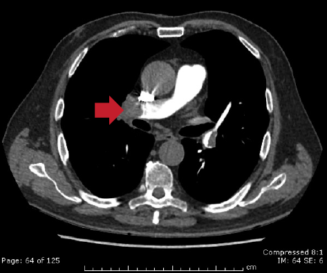
CT pulmonary angiography (CTPA) showinga bilateral pulmonary embolisms ...
Contrasted computed tomographic (CT) scan of the chest displaying a ...
Erythema around the sternal wound edges with surrounding blisters ...
Trametiglue provides durable inhibition of RAS/ERK signalling in models ...
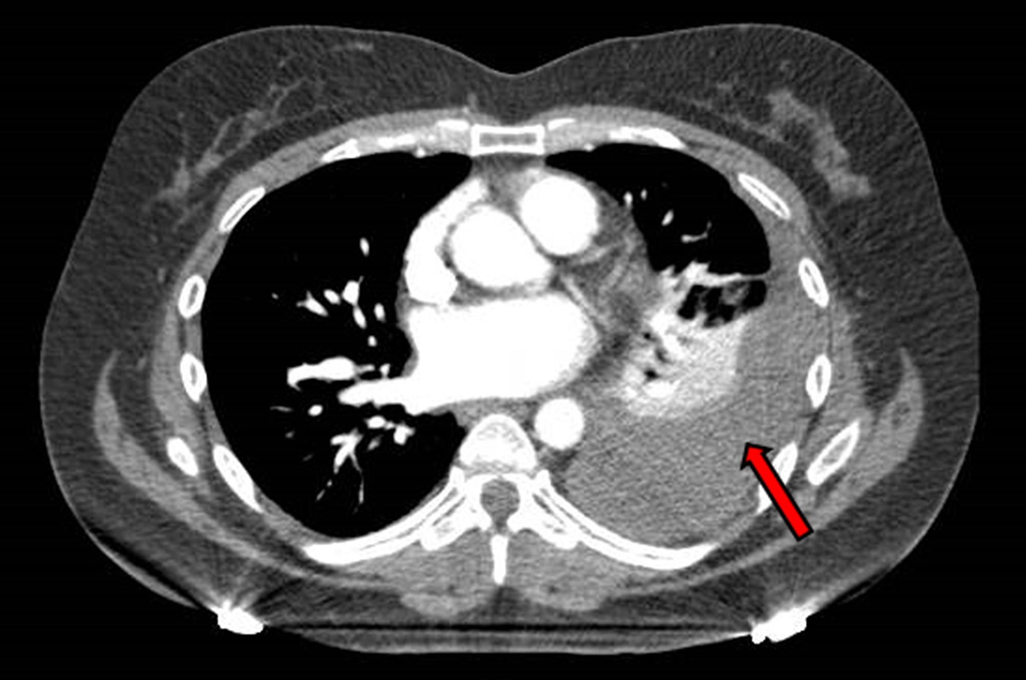
Refractory exudative pleural effusion in patients with chronic kidney ...
Troponin biomarkers: the benefits of echocardiography in a presumed ...
CT thorax showing left lower lobe lung cancer invading into inferior ...
Pulmonary infarction: What should I look for? | Eurorad
-CT neck with contrast demonstrates a mass compressing the esophagus at ...
Typical carcinoid occupying RUL main bronchus - CT scan Ca… | Flickr
Correction: An unusual presentation of pulmonary embolism leading to ...
CT chest with contrast (sagittal view) demonstrating catheter in the ...
The Heart of the Matter - The American Journal of Medicine
Chest CT in COVID-19: What the Radiologist Needs to Know | RadioGraphics
Respiratory Pathologies Flashcards | Quizlet
Article Fulle Text
Catastrophic Antiphospholipid Syndrome presenting as liver infarcts ...
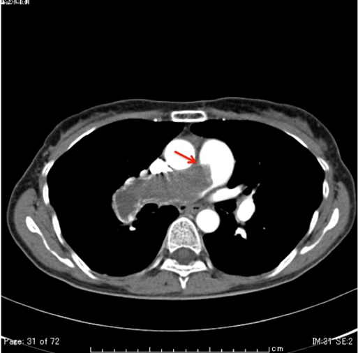
Computed tomography angiogram (CTA) with contrast demonstrates large ...
CT chest showing the left-sided pleural effusion. | Download Scientific ...
Pericardial tuberculosis | Eurorad
Cardiac computed tomography: thickening of parietal pericardium ...
Frontiers | Treating Heart Inflammation With Interleukin-1 Blockade in ...
Computed tomography of the thorax axial view with new mass-like ...
Spontaneous aortic intramural haematoma - with acute aortic syndrome ...
Patient 3: Axial image of CT pulmonary angiogram showing thrombi as ...
Figure 3 from Favorable course of pericardial angiosarcoma under ...
V1–V2–V3–V4 T wave inversion: left or right ventricle? | BMJ Case Reports
Chest CTA showed bilateral segmental pulmonary emboli, distal right ...
Follow-up CTA/aortogram three months following discharge showing total ...
CT angiography, axial image. The pulmonary trunk emerges from the right ...
pleural plaque
Multiple emboli caused by ascending aorta thrombus—Surgical approach ...