Please enter url.
Login
Logout
Please enter url.
Right Aortic Arch Ultrasound
mungfali.com
source
Comments
Fetal sonographic diagnosis of aortic arch anomalies - Yoo - 2003 ...
Fetal sonographic diagnosis of aortic arch anomalies - Yoo - 2003 ...
Fetal sonographic diagnosis of aortic arch anomalies - Yoo - 2003 ...
Prenatal ultrasonography and neonatal imaging of complete cleft sternum ...
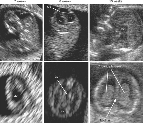
ULTRASOUND EVALUATION OF THE FETAL NEURAL AXIS | Radiology Key
Fetal sonographic diagnosis of aortic arch anomalies - Yoo - 2003 ...
Cardiac screening by STIC: can sonologists performing the 20‐week ...
An anatomical fetal brain structure and a normal variant mimicking ...
Fetal interrupted aortic arch: 2D‐4D echocardiography, associations and ...
(PDF) Transfontanelle ultrasonography of infant brain: Analysis of ...
Echocardiogram (Echo) on admission showing mild hypokinesis of left ...
Diagnosis and Resolution of Löeffler Endocarditis Secondary to ...
Heart | Radiology Key
of Fallot, Pulmonary Atresia with Ventricular Septal Defect, and Absent ...
Immagine correlata (With images) | Ultrasound, Sonography, Ultrasound humor
Prenatal Diagnosis of the Criss-Cross Heart - American Journal of ...
Echocardiographic characteristics and outcome of straddling mitral ...
ULTRASOUND EVALUATION OF THE FETAL HEART | Radiology Key
Echocardiography in the Patient with Right Heart Failure | Clinical Gate
38: Other Anomalies of the Ventricular Myocardium | Veterian Key
Initial echocardiogram in (A) 4-chamber view shows multiple segmental ...
Patient 2. Multiple cutaneous hemangiomas distributed over the face and ...
Placenta | Radiology Key
-Normal lungs, airways and diaphragm. (a) Prenatal ultrasound of the ...
Specimen of the anterior mitral valve leaflet with rupture of the ...
(PDF) Diagnosis of pericardial disease using percutaneous biopsy - Case ...
Practical Issues Related to the Examination, Anatomic Image Orientation ...
A) Pattern 1: sagittal scan of the fetal abdomen and thorax showing the ...
Abnormal or delayed development of the posterior membranous area of the ...
Preliminary Experience Using Motion Compensated CINE Magnetic Resonance ...
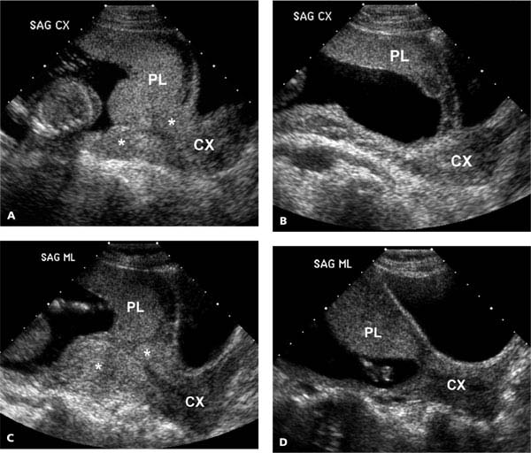
Ultrasound Evaluation of the Gravid Cervix | Obgyn Key
Anterior mitral leaflet tear. Note absence of calcification or ...
Fetal atrioventricular valve junction in normal fetuses and in fetuses ...
Echocardiographic images of cardiac rhabdomyomas located in (A) the LA ...
Fetal Diagnosis of Interrupted Aortic Arch - American Journal of Cardiology
Fetal-Right-Aortic-Arch
Aortic-Arch-Ultrasound
Aortic-Valve-Fetal-Ultrasound
Ductus-Venosus-Ultrasound
Double-Aortic-Arch-Ultrasound
Overriding-Aorta-Fetal-Ultrasound
Right-Sided-Aortic-Arch-Ultrasound
Bicuspid-Aortic-Valve-Fetal-Ultrasound
Interrupted-Aortic-Arch-Fetal-Ultrasound
Ductal-Arch-Fetal-Ultrasound
Fetal-Aortic-Stenosis
Aortic-Coarctation-Fetal-Ultrasound
Aortic-Arch-Embryology
Normal-Fetal-Heart-Ultrasound
Double-Aortic-Arch-Echocardiography
Interupted-Aortic-Arch-Ultrasound