Scanning electron microscope Stock Photos and Images
(4,262)See scanning electron microscope stock video clipsQuick filters:
Scanning electron microscope Stock Photos and Images
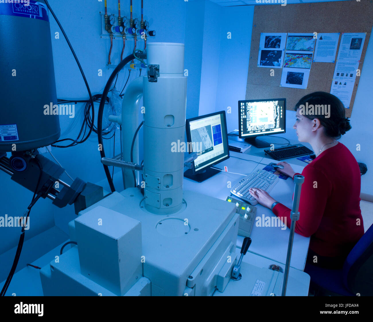 Scanning electron microscope in use at the Research Laboratory for Archaeology & the History of Art at the University of Oxford. Stock Photohttps://www.alamy.com/image-license-details/?v=1https://www.alamy.com/stock-photo-scanning-electron-microscope-in-use-at-the-research-laboratory-for-147196732.html
RMJFDAX4–Scanning electron microscope in use at the Research Laboratory for Archaeology & the History of Art at the University of Oxford.
 Researcher, Phillips FEI XL30 SFEG Scanning Electron Microscope. Stock Photohttps://www.alamy.com/image-license-details/?v=1https://www.alamy.com/stock-photo-researcher-phillips-fei-xl30-sfeg-scanning-electron-microscope-48209405.html
RMCPC3GD–Researcher, Phillips FEI XL30 SFEG Scanning Electron Microscope.
 A dual computer monitor display showing the software controlling a JEOL scanning electron microscope and the image it is seeing. Stock Photohttps://www.alamy.com/image-license-details/?v=1https://www.alamy.com/stock-photo-a-dual-computer-monitor-display-showing-the-software-controlling-a-72522487.html
RME5YK4R–A dual computer monitor display showing the software controlling a JEOL scanning electron microscope and the image it is seeing.
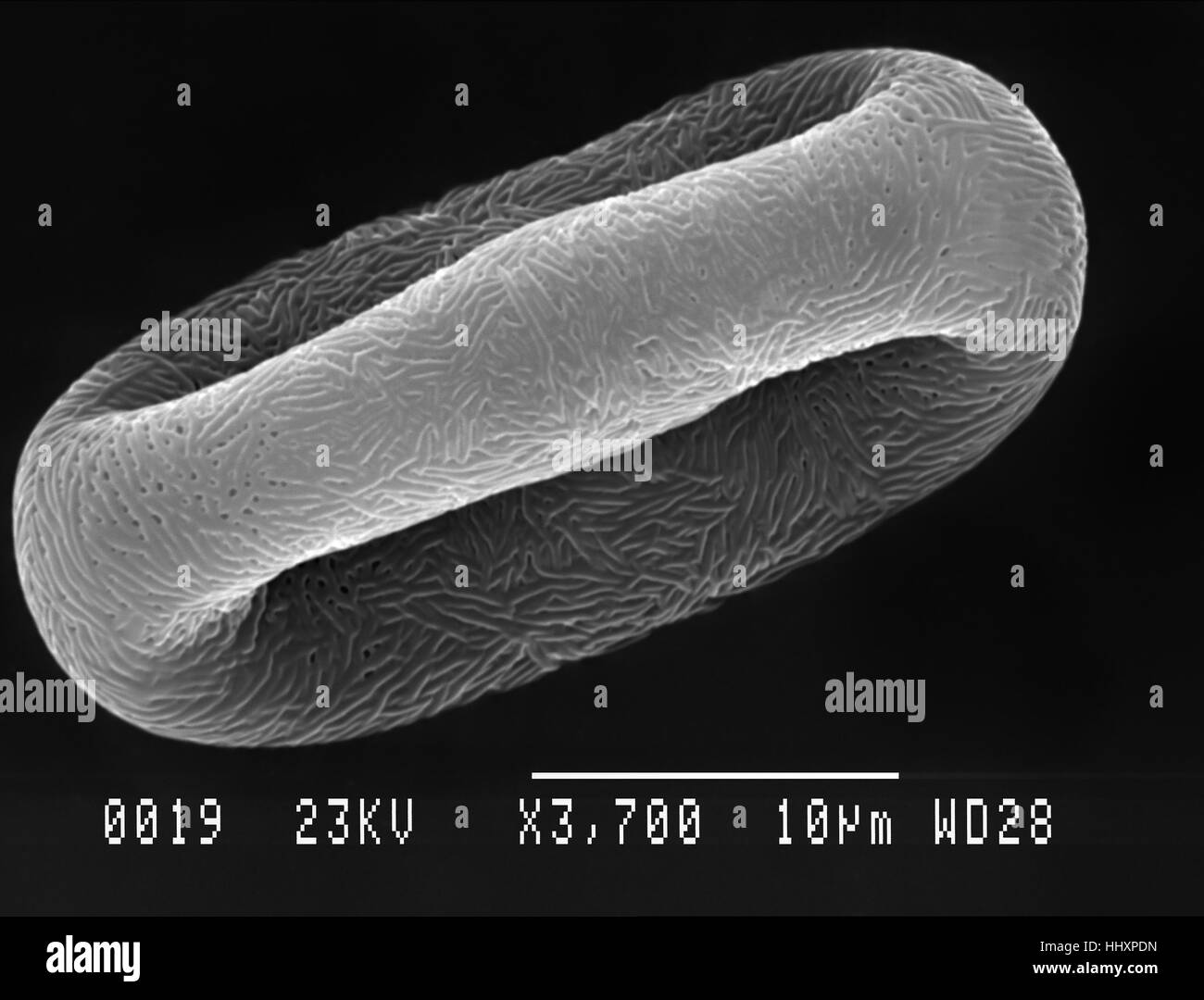 Cow parsley (Anthriscus sylvestris) pollen particle magnified under scanning electron microscope (SEM) Stock Photohttps://www.alamy.com/image-license-details/?v=1https://www.alamy.com/stock-photo-cow-parsley-anthriscus-sylvestris-pollen-particle-magnified-under-131510113.html
RFHHXPDN–Cow parsley (Anthriscus sylvestris) pollen particle magnified under scanning electron microscope (SEM)
 Tiny fungus gnat under scanning electron microscope Stock Photohttps://www.alamy.com/image-license-details/?v=1https://www.alamy.com/tiny-fungus-gnat-under-scanning-electron-microscope-image214877716.html
RMPDGEM4–Tiny fungus gnat under scanning electron microscope
 Optical electron scanning microscope concept art, 3D illustration Stock Photohttps://www.alamy.com/image-license-details/?v=1https://www.alamy.com/stock-image-optical-electron-scanning-microscope-concept-art-3d-illustration-164842240.html
RFKG55XT–Optical electron scanning microscope concept art, 3D illustration
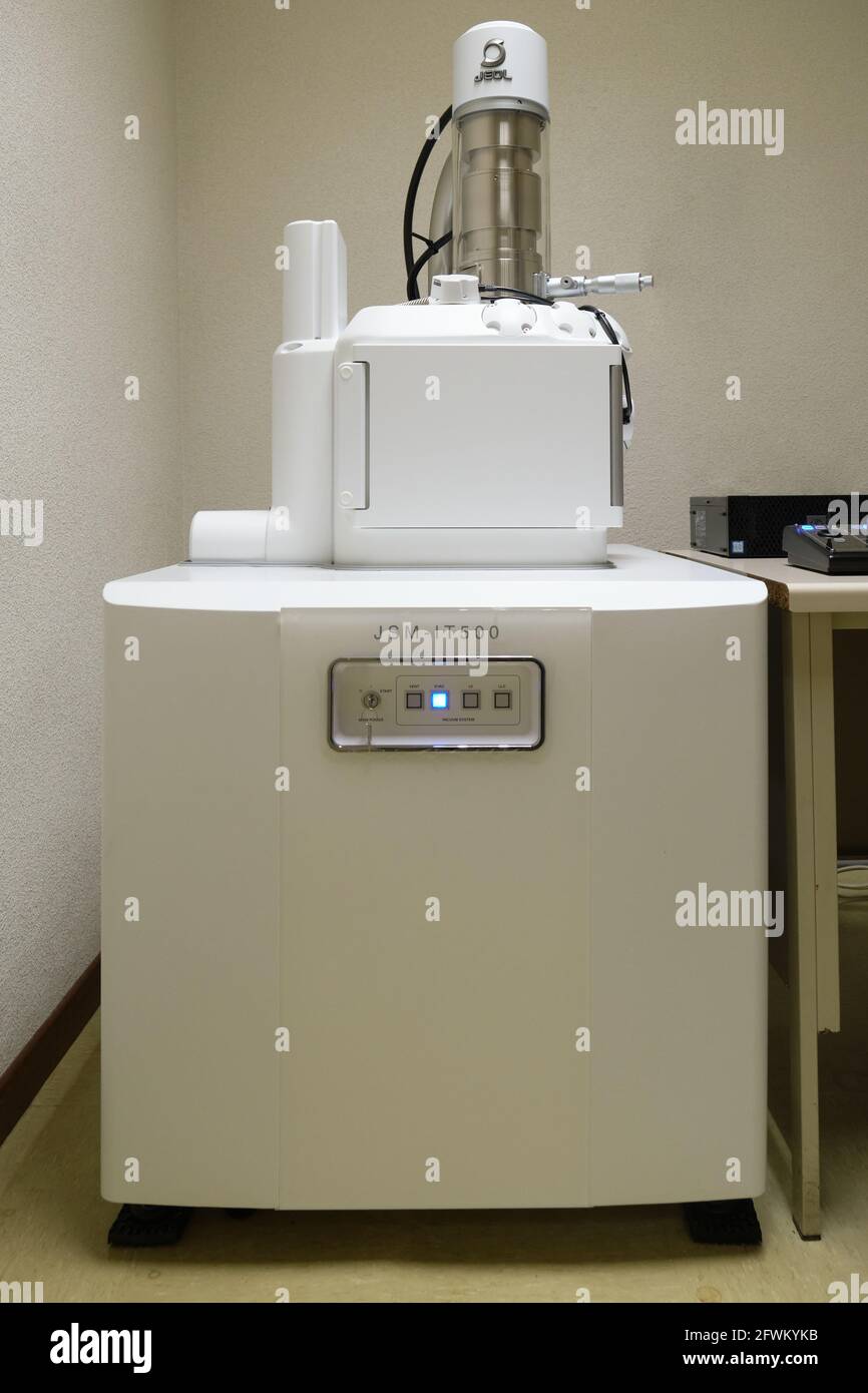 Madrid, Spain. May 12, 2021: JSM-IT500 InTouchScope™ Scanning Electron Microscope Stock Photohttps://www.alamy.com/image-license-details/?v=1https://www.alamy.com/madrid-spain-may-12-2021-jsm-it500-intouchscope-scanning-electron-microscope-image428854031.html
RF2FWKYKB–Madrid, Spain. May 12, 2021: JSM-IT500 InTouchScope™ Scanning Electron Microscope
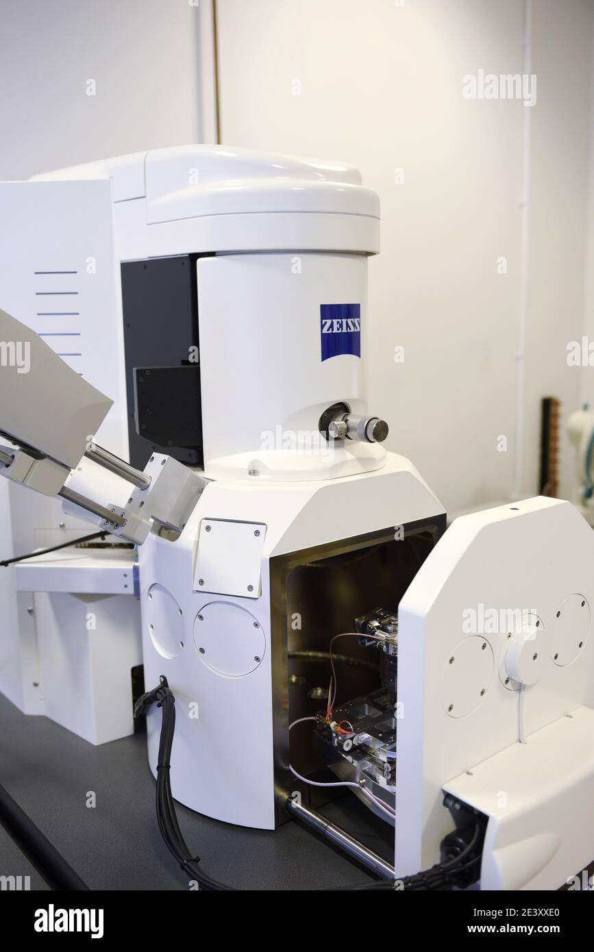 Zeiss EVO 15 Scanning Electron Microscope scanning station in a science lab. It is flexible variable pressure scanning electron microscope or SEM with Stock Photohttps://www.alamy.com/image-license-details/?v=1https://www.alamy.com/zeiss-evo-15-scanning-electron-microscope-scanning-station-in-a-science-lab-it-is-flexible-variable-pressure-scanning-electron-microscope-or-sem-with-image398273960.html
RM2E3XXE0–Zeiss EVO 15 Scanning Electron Microscope scanning station in a science lab. It is flexible variable pressure scanning electron microscope or SEM with
 Cast Iron fractured surface. Fatigue striations under iron oxide corrosion imaged in a scanning electron microscope Stock Photohttps://www.alamy.com/image-license-details/?v=1https://www.alamy.com/stock-photo-cast-iron-fractured-surface-fatigue-striations-under-iron-oxide-corrosion-86281113.html
RFF0ACC9–Cast Iron fractured surface. Fatigue striations under iron oxide corrosion imaged in a scanning electron microscope
 Scientist sitting in analytical laboratory with scanning electron microscope and spectrometer Stock Photohttps://www.alamy.com/image-license-details/?v=1https://www.alamy.com/scientist-sitting-in-analytical-laboratory-with-scanning-electron-image69861376.html
RFE1JCW4–Scientist sitting in analytical laboratory with scanning electron microscope and spectrometer
 Red Blood Cells.Scanning electron microscope Stock Photohttps://www.alamy.com/image-license-details/?v=1https://www.alamy.com/red-blood-cellsscanning-electron-microscope-image209285722.html
RFP4DP22–Red Blood Cells.Scanning electron microscope
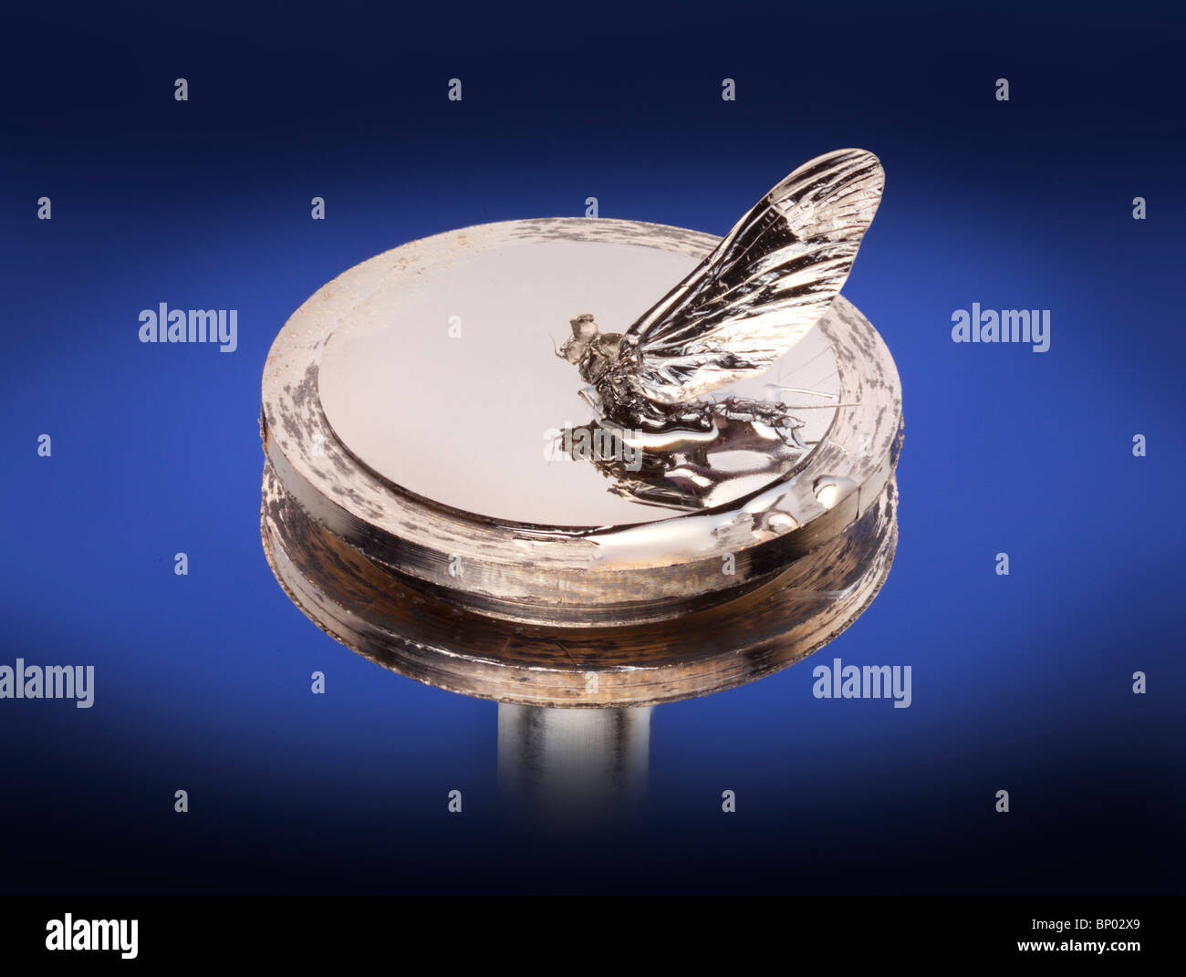 S.E.M. specimen support stub, scanning electron microscope, coated specimen Stock Photohttps://www.alamy.com/image-license-details/?v=1https://www.alamy.com/stock-photo-sem-specimen-support-stub-scanning-electron-microscope-coated-specimen-30735105.html
RMBP02X9–S.E.M. specimen support stub, scanning electron microscope, coated specimen
 Dobsonfly (Megaloptera: Corydalidae) imaged in a scanning electron microscope Stock Photohttps://www.alamy.com/image-license-details/?v=1https://www.alamy.com/stock-photo-dobsonfly-megaloptera-corydalidae-imaged-in-a-scanning-electron-microscope-140114286.html
RFJ3XN5J–Dobsonfly (Megaloptera: Corydalidae) imaged in a scanning electron microscope
 colorized scanning electron microscope image of a blood clot Stock Photohttps://www.alamy.com/image-license-details/?v=1https://www.alamy.com/stock-photo-colorized-scanning-electron-microscope-image-of-a-blood-clot-17533198.html
RMB0EKNJ–colorized scanning electron microscope image of a blood clot
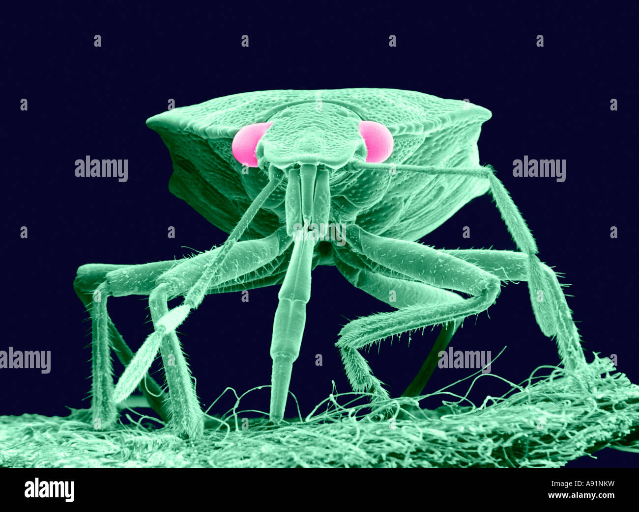 Scanning Electron Microscope image of a Plant Bug magnified approximately 30X (Color enhanced) Stock Photohttps://www.alamy.com/image-license-details/?v=1https://www.alamy.com/stock-photo-scanning-electron-microscope-image-of-a-plant-bug-magnified-approximately-12222012.html
RFA91NKW–Scanning Electron Microscope image of a Plant Bug magnified approximately 30X (Color enhanced)
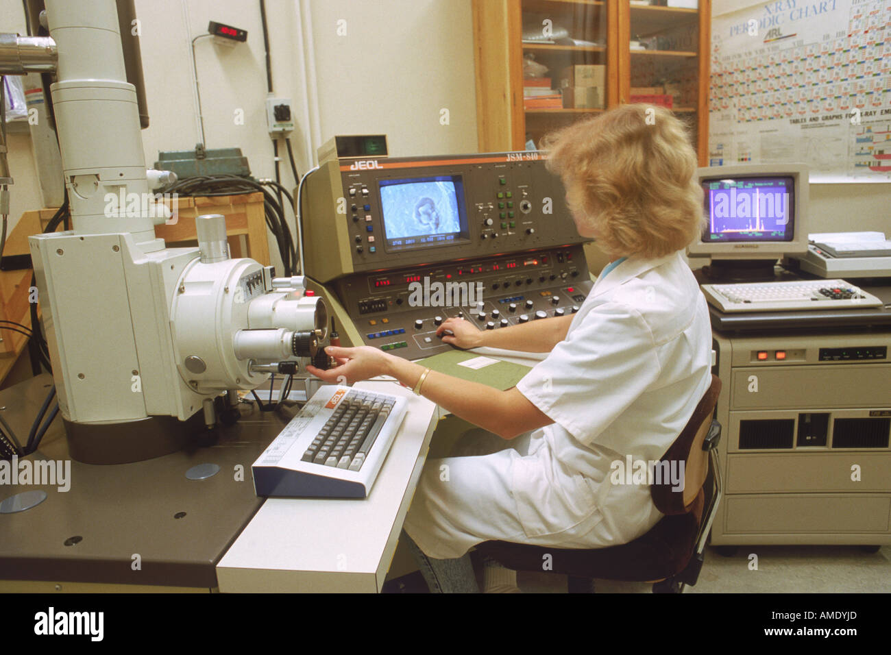 Woman in research lab with scanning electron microscope and x ray analyzer Stock Photohttps://www.alamy.com/image-license-details/?v=1https://www.alamy.com/woman-in-research-lab-with-scanning-electron-microscope-and-x-ray-image8705388.html
RMAMDYJD–Woman in research lab with scanning electron microscope and x ray analyzer
 Researchers at the Texas Center for Cancer Nanomedicine (TCCN) have created particle-based vaccines for the treatment of cancer. The particles carry molecules that stimulate immune cells and cancer antigens (proteins) that direct the immune response. This scanning electron microscope image shows dendritic cells, pseudostained green, interacting with T cells, pseudostained pink. Dendritic cells internalize particles, provoke antigens and present peptides to T cells to direct immune responses. Stock Photohttps://www.alamy.com/image-license-details/?v=1https://www.alamy.com/researchers-at-the-texas-center-for-cancer-nanomedicine-tccn-have-created-particle-based-vaccines-for-the-treatment-of-cancer-the-particles-carry-molecules-that-stimulate-immune-cells-and-cancer-antigens-proteins-that-direct-the-immune-response-this-scanning-electron-microscope-image-shows-dendritic-cells-pseudostained-green-interacting-with-t-cells-pseudostained-pink-dendritic-cells-internalize-particles-provoke-antigens-and-present-peptides-to-t-cells-to-direct-immune-responses-image476707123.html
RM2JKFTPB–Researchers at the Texas Center for Cancer Nanomedicine (TCCN) have created particle-based vaccines for the treatment of cancer. The particles carry molecules that stimulate immune cells and cancer antigens (proteins) that direct the immune response. This scanning electron microscope image shows dendritic cells, pseudostained green, interacting with T cells, pseudostained pink. Dendritic cells internalize particles, provoke antigens and present peptides to T cells to direct immune responses.
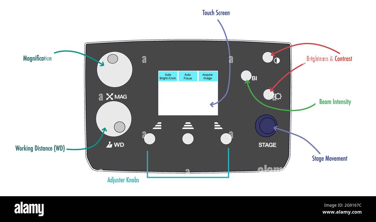 Scanning electron microscope control panel, illustration Stock Photohttps://www.alamy.com/image-license-details/?v=1https://www.alamy.com/scanning-electron-microscope-control-panel-illustration-image435817968.html
RF2G9167C–Scanning electron microscope control panel, illustration
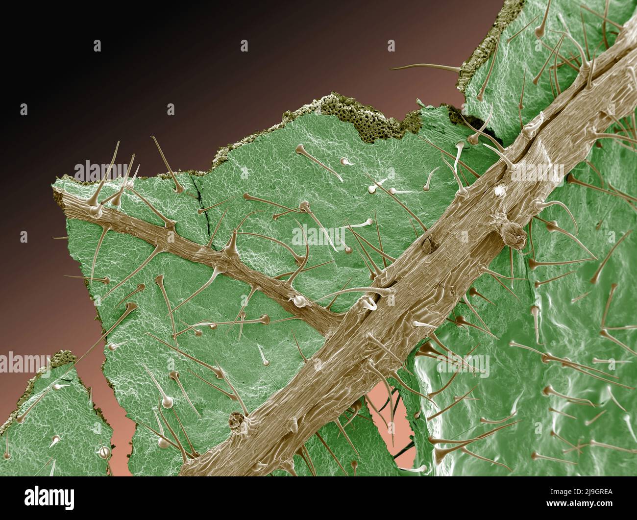 SEM Scanning Electron Microscope image of stinging nettles Stock Photohttps://www.alamy.com/image-license-details/?v=1https://www.alamy.com/sem-scanning-electron-microscope-image-of-stinging-nettles-image470581506.html
RM2J9GREA–SEM Scanning Electron Microscope image of stinging nettles
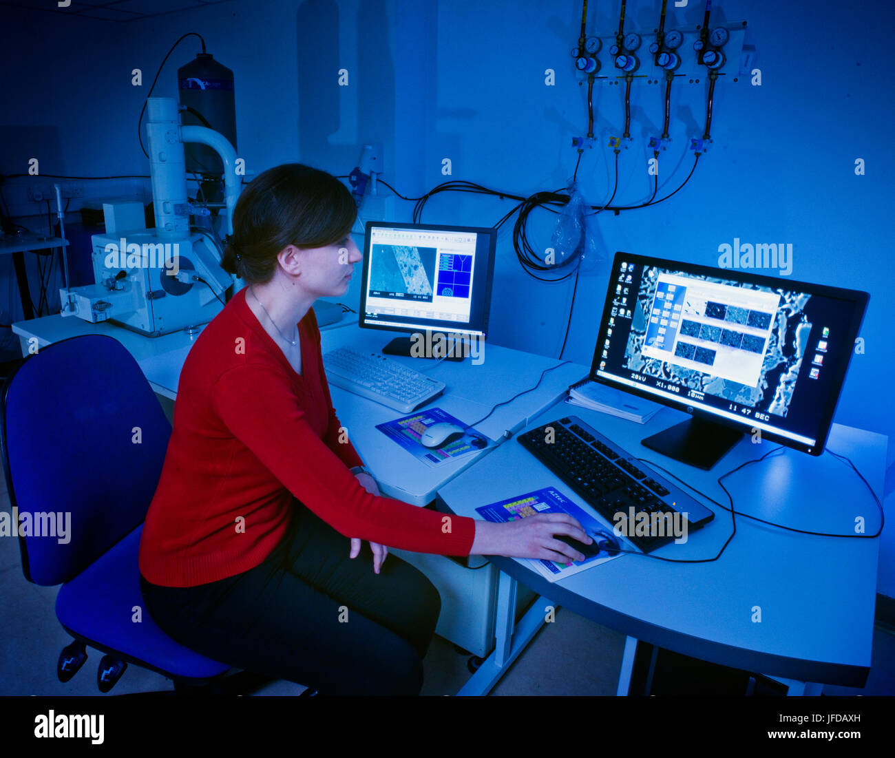 Scanning electron microscope in use at the Research Laboratory for Archaeology & the History of Art at the University of Oxford. Stock Photohttps://www.alamy.com/image-license-details/?v=1https://www.alamy.com/stock-photo-scanning-electron-microscope-in-use-at-the-research-laboratory-for-147196745.html
RMJFDAXH–Scanning electron microscope in use at the Research Laboratory for Archaeology & the History of Art at the University of Oxford.
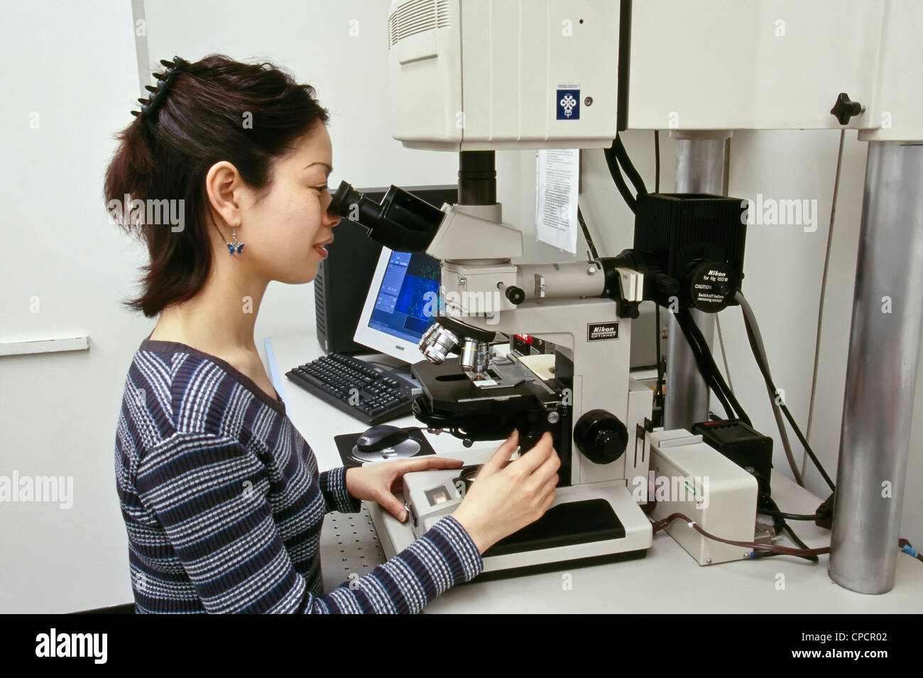 Researcher, Nikon MRC1024 Confocal/UV Laser Scanning Microscope. Stock Photohttps://www.alamy.com/image-license-details/?v=1https://www.alamy.com/stock-photo-researcher-nikon-mrc1024-confocaluv-laser-scanning-microscope-48224626.html
RMCPCR02–Researcher, Nikon MRC1024 Confocal/UV Laser Scanning Microscope.
 Scanning electron micrograph of HIV particles infecting a human H9 T cell. Stock Photohttps://www.alamy.com/image-license-details/?v=1https://www.alamy.com/stock-photo-scanning-electron-micrograph-of-hiv-particles-infecting-a-human-h9-76547999.html
RFECF1N3–Scanning electron micrograph of HIV particles infecting a human H9 T cell.
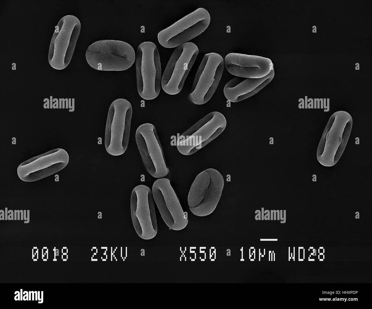 Many cow parsley (Anthriscus sylvestris) pollen grains magnified under scanning electron microscope (SEM), demonstrating shape at different angles. Stock Photohttps://www.alamy.com/image-license-details/?v=1https://www.alamy.com/stock-photo-many-cow-parsley-anthriscus-sylvestris-pollen-grains-magnified-under-131510114.html
RFHHXPDP–Many cow parsley (Anthriscus sylvestris) pollen grains magnified under scanning electron microscope (SEM), demonstrating shape at different angles.
 Scanning electron microscope with EMF microanalysis Stock Photohttps://www.alamy.com/image-license-details/?v=1https://www.alamy.com/scanning-electron-microscope-with-emf-microanalysis-image576300719.html
RF2TDGNCF–Scanning electron microscope with EMF microanalysis
 Optical electron scanning microscope virus research concept Stock Photohttps://www.alamy.com/image-license-details/?v=1https://www.alamy.com/stock-image-optical-electron-scanning-microscope-virus-research-concept-164842243.html
RFKG55XY–Optical electron scanning microscope virus research concept
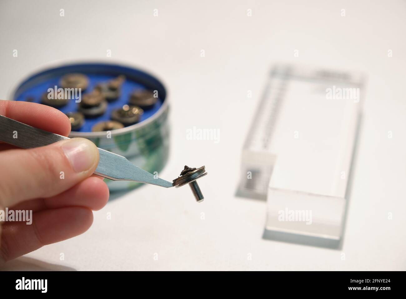 Scientific hand holding a tweezers with a scanning electron microscope sample on a specimen mount. SEM pins to analyze. Stock Photohttps://www.alamy.com/image-license-details/?v=1https://www.alamy.com/scientific-hand-holding-a-tweezers-with-a-scanning-electron-microscope-sample-on-a-specimen-mount-sem-pins-to-analyze-image426560348.html
RF2FNYE24–Scientific hand holding a tweezers with a scanning electron microscope sample on a specimen mount. SEM pins to analyze.
 Zeiss EVO 15 Scanning Electron Microscope scanning station in a science lab. It is flexible variable pressure scanning electron microscope or SEM with Stock Photohttps://www.alamy.com/image-license-details/?v=1https://www.alamy.com/zeiss-evo-15-scanning-electron-microscope-scanning-station-in-a-science-lab-it-is-flexible-variable-pressure-scanning-electron-microscope-or-sem-with-image398273964.html
RM2E3XXE4–Zeiss EVO 15 Scanning Electron Microscope scanning station in a science lab. It is flexible variable pressure scanning electron microscope or SEM with
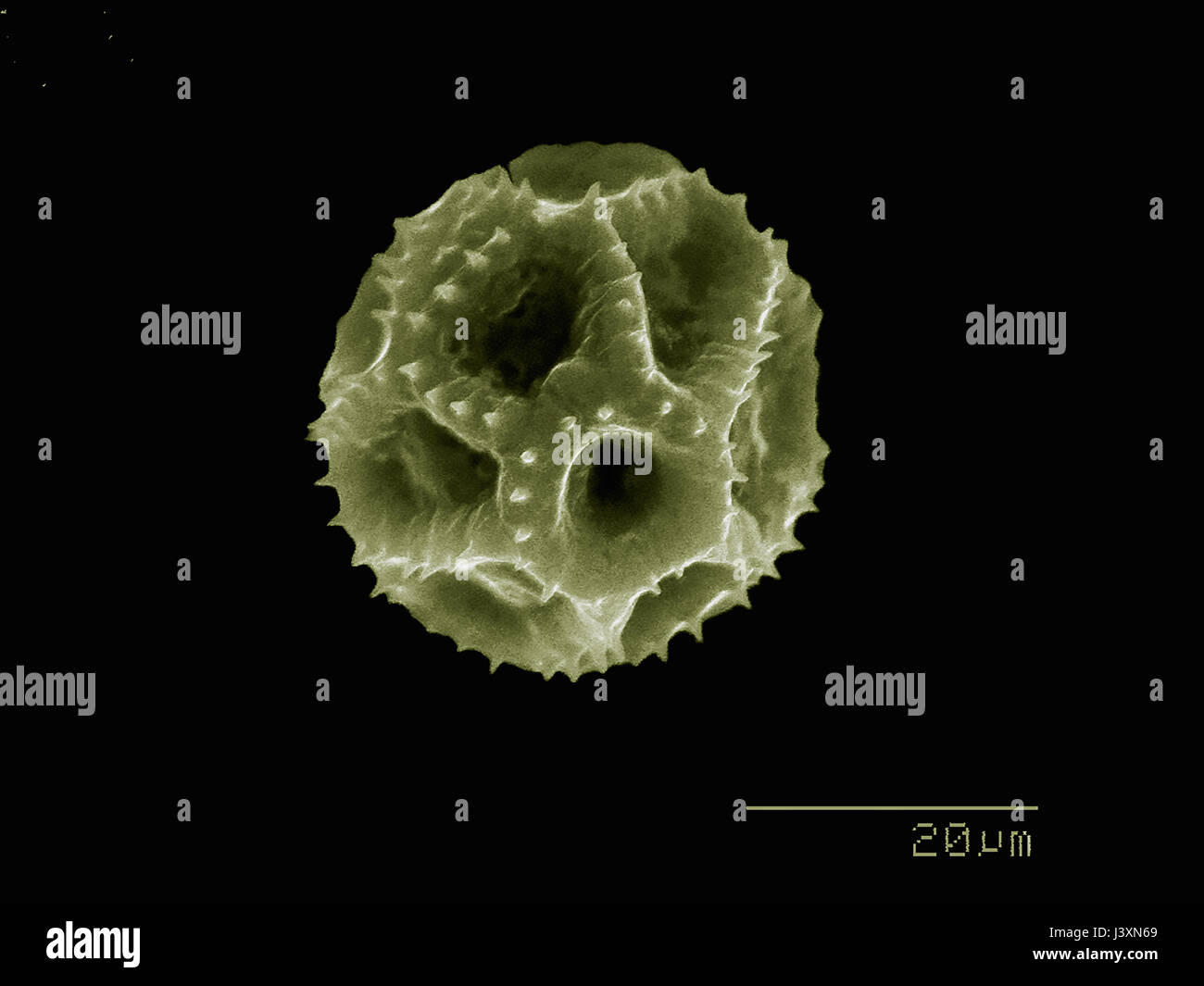 Dandelion pollen imaged in a scanning electron microscope Stock Photohttps://www.alamy.com/image-license-details/?v=1https://www.alamy.com/stock-photo-dandelion-pollen-imaged-in-a-scanning-electron-microscope-140114305.html
RFJ3XN69–Dandelion pollen imaged in a scanning electron microscope
 Scientist standing in analytical laboratory with scanning electron microscope and spectrometer Stock Photohttps://www.alamy.com/image-license-details/?v=1https://www.alamy.com/scientist-standing-in-analytical-laboratory-with-scanning-electron-image69861379.html
RFE1JCW7–Scientist standing in analytical laboratory with scanning electron microscope and spectrometer
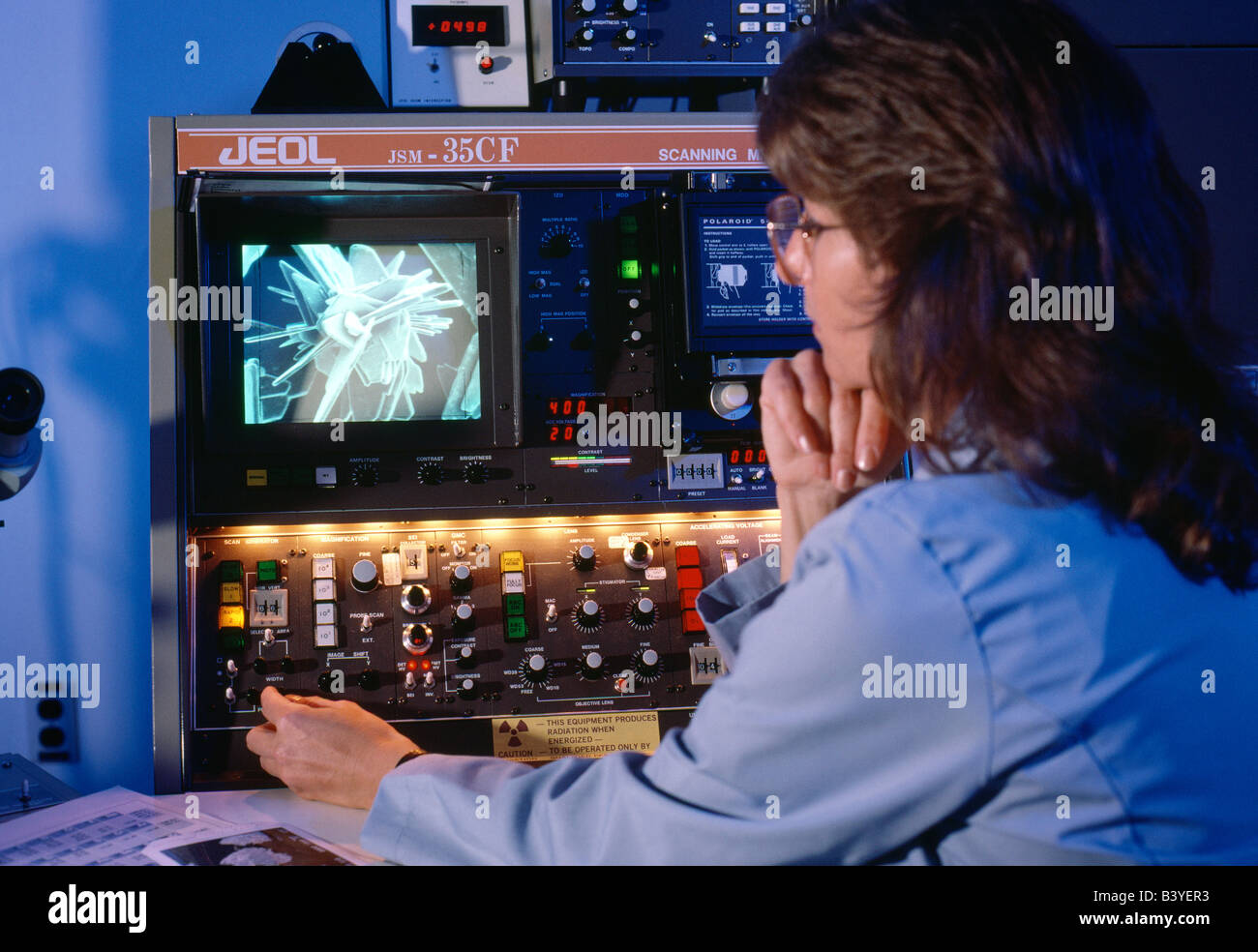 Female scientist uses scanning electron microscope to analyze materials Stock Photohttps://www.alamy.com/image-license-details/?v=1https://www.alamy.com/stock-photo-female-scientist-uses-scanning-electron-microscope-to-analyze-materials-19658663.html
RMB3YER3–Female scientist uses scanning electron microscope to analyze materials
 Scanning electron microscope microscope in a physical lab Stock Photohttps://www.alamy.com/image-license-details/?v=1https://www.alamy.com/scanning-electron-microscope-microscope-in-a-physical-lab-image327935921.html
RF2A1ENH5–Scanning electron microscope microscope in a physical lab
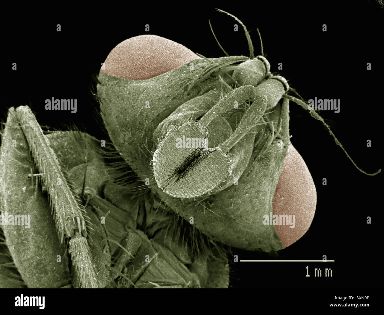 Head of house fly (Muscidae) imaged in a scanning electron microscope Stock Photohttps://www.alamy.com/image-license-details/?v=1https://www.alamy.com/stock-photo-head-of-house-fly-muscidae-imaged-in-a-scanning-electron-microscope-140114402.html
RFJ3XN9P–Head of house fly (Muscidae) imaged in a scanning electron microscope
 Scanning electron microscope (SEM) image of a human macrophage Stock Photohttps://www.alamy.com/image-license-details/?v=1https://www.alamy.com/stock-photo-scanning-electron-microscope-sem-image-of-a-human-macrophage-26900632.html
RMBFNC0T–Scanning electron microscope (SEM) image of a human macrophage
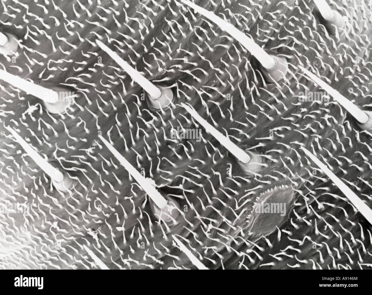 Scanning Electron Microscope close-up of the hairs on the legs of a plant bug magnified approximately 800X Stock Photohttps://www.alamy.com/image-license-details/?v=1https://www.alamy.com/stock-photo-scanning-electron-microscope-close-up-of-the-hairs-on-the-legs-of-12216139.html
RFA9146M–Scanning Electron Microscope close-up of the hairs on the legs of a plant bug magnified approximately 800X
 GREAT BRITAIN - CIRCA 1994: A used postage stamp from the UK, commemorating the invention of the Scanning Electron Microscope, circa 1994. Stock Photohttps://www.alamy.com/image-license-details/?v=1https://www.alamy.com/stock-photo-great-britain-circa-1994-a-used-postage-stamp-from-the-uk-commemorating-131814332.html
RMHJCJEM–GREAT BRITAIN - CIRCA 1994: A used postage stamp from the UK, commemorating the invention of the Scanning Electron Microscope, circa 1994.
 Scanning electron microscope image of regulatory T cells (red) interacting with antigen-presenting cells (blue). T regulatory cells can suppress T cell responses to maintain homeostasis in the immune system. Stock Photohttps://www.alamy.com/image-license-details/?v=1https://www.alamy.com/scanning-electron-microscope-image-of-regulatory-t-cells-red-interacting-with-antigen-presenting-cells-blue-t-regulatory-cells-can-suppress-t-cell-responses-to-maintain-homeostasis-in-the-immune-system-image476707127.html
RM2JKFTPF–Scanning electron microscope image of regulatory T cells (red) interacting with antigen-presenting cells (blue). T regulatory cells can suppress T cell responses to maintain homeostasis in the immune system.
 Surgical needle, SEM Stock Photohttps://www.alamy.com/image-license-details/?v=1https://www.alamy.com/surgical-needle-sem-image341959471.html
RF2AT9GP7–Surgical needle, SEM
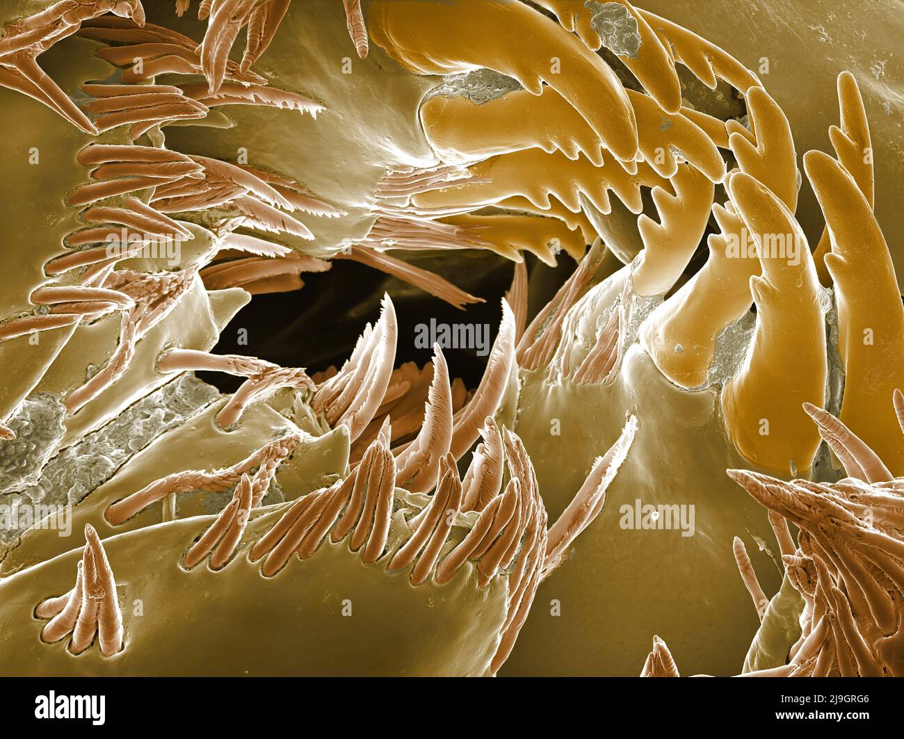 SEM Scanning Electron Microscope image of a Sandhopper, Sand Flea, amphipod Stock Photohttps://www.alamy.com/image-license-details/?v=1https://www.alamy.com/sem-scanning-electron-microscope-image-of-a-sandhopper-sand-flea-amphipod-image470581558.html
RM2J9GRG6–SEM Scanning Electron Microscope image of a Sandhopper, Sand Flea, amphipod
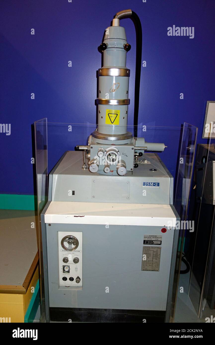 a scanning electron microscope Stock Photohttps://www.alamy.com/image-license-details/?v=1https://www.alamy.com/a-scanning-electron-microscope-image373157326.html
RF2CK2NYA–a scanning electron microscope
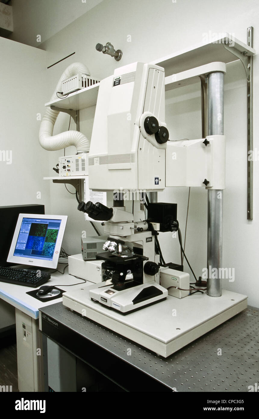 Nikon MRC1024 Confocal/UV Laser Scanning Microscope. Stock Photohttps://www.alamy.com/image-license-details/?v=1https://www.alamy.com/stock-photo-nikon-mrc1024-confocaluv-laser-scanning-microscope-48209397.html
RMCPC3G5–Nikon MRC1024 Confocal/UV Laser Scanning Microscope.
 Colorized scanning electron micrograph of a T lymphocyte. Stock Photohttps://www.alamy.com/image-license-details/?v=1https://www.alamy.com/stock-photo-colorized-scanning-electron-micrograph-of-a-t-lymphocyte-130443046.html
RFHG65C6–Colorized scanning electron micrograph of a T lymphocyte.
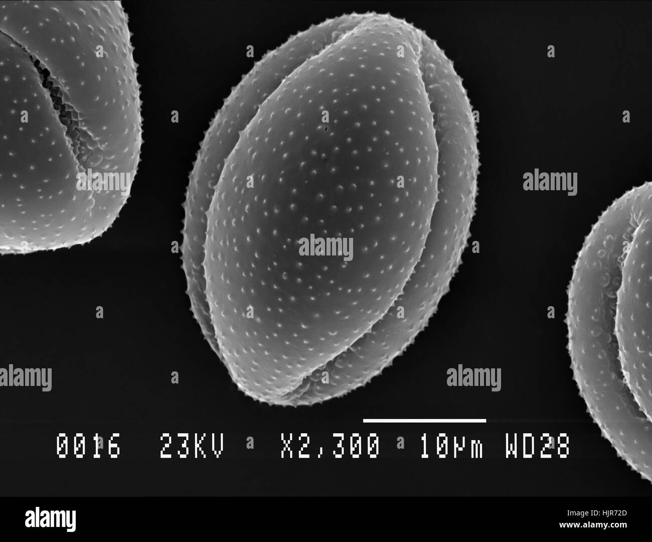 Scanning electron micrograph of one pollen particle from Lesser Celandine flower. Nottingham, UK. April. Stock Photohttps://www.alamy.com/image-license-details/?v=1https://www.alamy.com/stock-photo-scanning-electron-micrograph-of-one-pollen-particle-from-lesser-celandine-132046837.html
RFHJR72D–Scanning electron micrograph of one pollen particle from Lesser Celandine flower. Nottingham, UK. April.
 Scanning electron microscope with EMF microanalysis Stock Photohttps://www.alamy.com/image-license-details/?v=1https://www.alamy.com/scanning-electron-microscope-with-emf-microanalysis-image608624461.html
RF2XA56KW–Scanning electron microscope with EMF microanalysis
 Matal workpiece under the electron microscope Stock Photohttps://www.alamy.com/image-license-details/?v=1https://www.alamy.com/matal-workpiece-under-the-electron-microscope-image566626757.html
RF2RWT25W–Matal workpiece under the electron microscope
 Scientific hand holding a tweezers with a scanning electron microscope sample on a specimen mount. SEM pins to analyze. Stock Photohttps://www.alamy.com/image-license-details/?v=1https://www.alamy.com/scientific-hand-holding-a-tweezers-with-a-scanning-electron-microscope-sample-on-a-specimen-mount-sem-pins-to-analyze-image428854025.html
RF2FWKYK5–Scientific hand holding a tweezers with a scanning electron microscope sample on a specimen mount. SEM pins to analyze.
 Zeiss EVO 15 Scanning Electron Microscope scanning station in a science lab. It is flexible variable pressure scanning electron microscope or SEM with Stock Photohttps://www.alamy.com/image-license-details/?v=1https://www.alamy.com/zeiss-evo-15-scanning-electron-microscope-scanning-station-in-a-science-lab-it-is-flexible-variable-pressure-scanning-electron-microscope-or-sem-with-image398273968.html
RM2E3XXE8–Zeiss EVO 15 Scanning Electron Microscope scanning station in a science lab. It is flexible variable pressure scanning electron microscope or SEM with
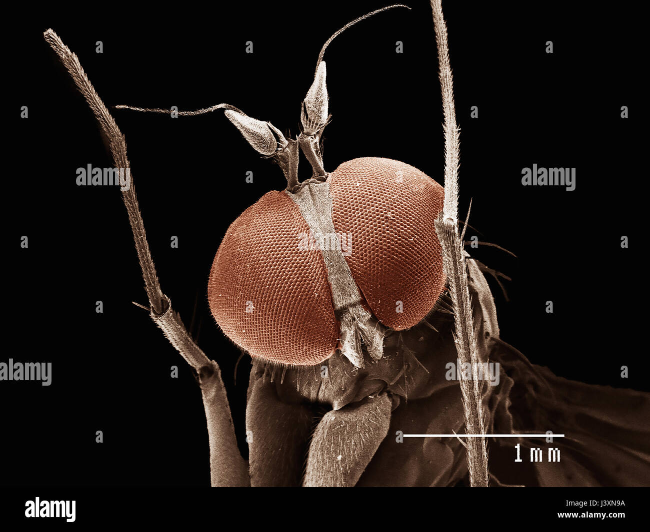 Head of long legged fly (dolichopodiae) imaged in a scanning electron microscope Stock Photohttps://www.alamy.com/image-license-details/?v=1https://www.alamy.com/stock-photo-head-of-long-legged-fly-dolichopodiae-imaged-in-a-scanning-electron-140114390.html
RFJ3XN9A–Head of long legged fly (dolichopodiae) imaged in a scanning electron microscope
 Scientist standing in analytical laboratory with scanning electron microscope and spectrometer Stock Photohttps://www.alamy.com/image-license-details/?v=1https://www.alamy.com/scientist-standing-in-analytical-laboratory-with-scanning-electron-image69861384.html
RFE1JCWC–Scientist standing in analytical laboratory with scanning electron microscope and spectrometer
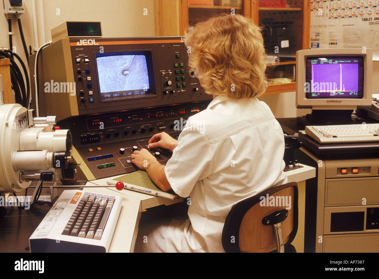 Woman in research lab with scanning electron microscope and x ray analyzer Stock Photohttps://www.alamy.com/image-license-details/?v=1https://www.alamy.com/woman-in-research-lab-with-scanning-electron-microscope-and-x-ray-image1471366.html
RFAP7387–Woman in research lab with scanning electron microscope and x ray analyzer
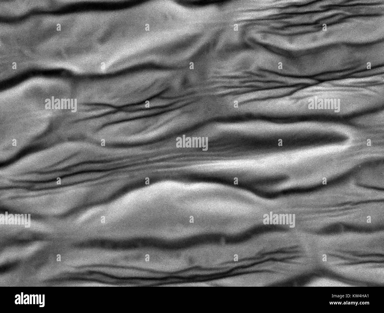 Scanning electron microscope (SEM) micrograph depicting the petal of a carnation flower (Dianthus caryophyllus), showing cellular structure, with creasing due to desication (drying) of the petal, at a magnification of 1500x, 2016. Stock Photohttps://www.alamy.com/image-license-details/?v=1https://www.alamy.com/stock-photo-scanning-electron-microscope-sem-micrograph-depicting-the-petal-of-170361129.html
RMKW4HA1–Scanning electron microscope (SEM) micrograph depicting the petal of a carnation flower (Dianthus caryophyllus), showing cellular structure, with creasing due to desication (drying) of the petal, at a magnification of 1500x, 2016.
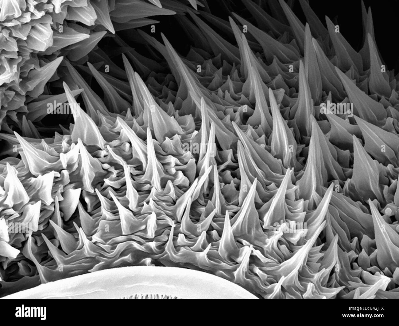 Large Caterpillar: Gold coated and imaged in Scanning electron microscope Stock Photohttps://www.alamy.com/image-license-details/?v=1https://www.alamy.com/stock-photo-large-caterpillar-gold-coated-and-imaged-in-scanning-electron-microscope-71358810.html
RFE42JTX–Large Caterpillar: Gold coated and imaged in Scanning electron microscope
 Scanning electron microscope (SEM) image of a human macrophage Stock Photohttps://www.alamy.com/image-license-details/?v=1https://www.alamy.com/stock-photo-scanning-electron-microscope-sem-image-of-a-human-macrophage-26900649.html
RMBFNC1D–Scanning electron microscope (SEM) image of a human macrophage
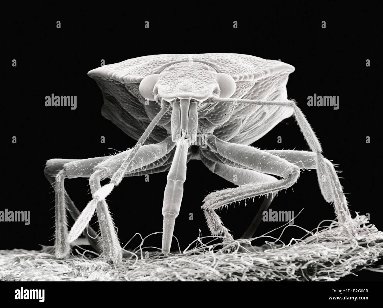 Scanning electron microscope close-up image of a beetle Stock Photohttps://www.alamy.com/image-license-details/?v=1https://www.alamy.com/stock-photo-scanning-electron-microscope-close-up-image-of-a-beetle-18790935.html
RFB2G00R–Scanning electron microscope close-up image of a beetle
 Optical electron scanning microscope laser beam. Concept art 3D illustration Stock Photohttps://www.alamy.com/image-license-details/?v=1https://www.alamy.com/stock-image-optical-electron-scanning-microscope-laser-beam-concept-art-3d-illustration-164842237.html
RFKG55XN–Optical electron scanning microscope laser beam. Concept art 3D illustration
 This scanning electron microscope image shows SARS-CoV-2 (round orange particles) emerging from the surface of a cell cultured in the laboratory. SARS-CoV-2, also known as 2019-nCoV, is the virus that causes COVID-19. Image captured and colorized at Rocky Mountain Laboratories in Hamilton, Montana. Credit: NIAID Stock Photohttps://www.alamy.com/image-license-details/?v=1https://www.alamy.com/this-scanning-electron-microscope-image-shows-sars-cov-2-round-orange-particles-emerging-from-the-surface-of-a-cell-cultured-in-the-laboratory-sars-cov-2-also-known-as-2019-ncov-is-the-virus-that-causes-covid-19-image-captured-and-colorized-at-rocky-mountain-laboratories-in-hamilton-montana-credit-niaid-image476706638.html
RM2JKFT52–This scanning electron microscope image shows SARS-CoV-2 (round orange particles) emerging from the surface of a cell cultured in the laboratory. SARS-CoV-2, also known as 2019-nCoV, is the virus that causes COVID-19. Image captured and colorized at Rocky Mountain Laboratories in Hamilton, Montana. Credit: NIAID
 Surgical needle, SEM Stock Photohttps://www.alamy.com/image-license-details/?v=1https://www.alamy.com/surgical-needle-sem-image341959514.html
RF2AT9GRP–Surgical needle, SEM
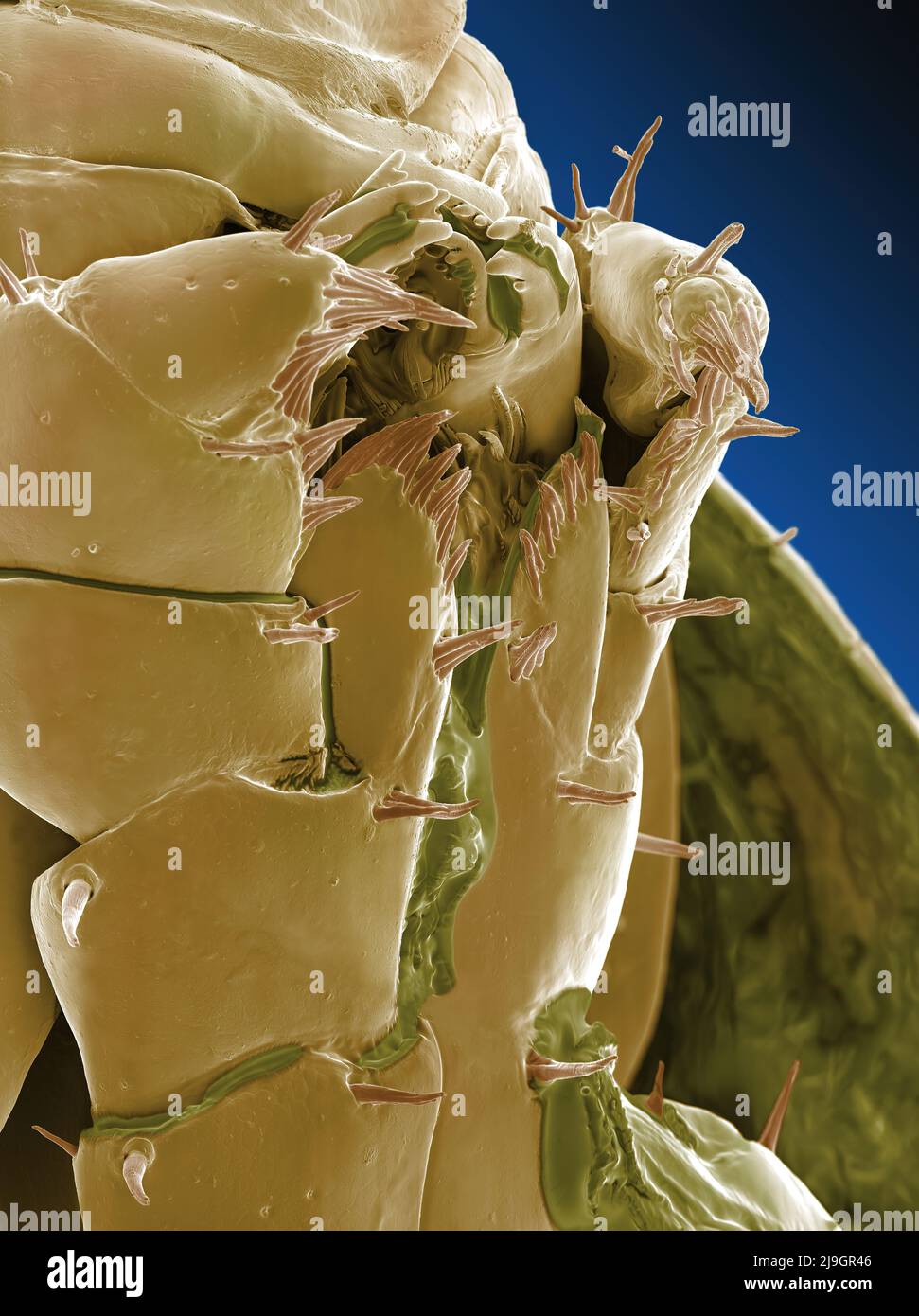 SEM Scanning Electron Microscope image of a Sandhopper, Sand Flea, amphipod Stock Photohttps://www.alamy.com/image-license-details/?v=1https://www.alamy.com/sem-scanning-electron-microscope-image-of-a-sandhopper-sand-flea-amphipod-image470581222.html
RM2J9GR46–SEM Scanning Electron Microscope image of a Sandhopper, Sand Flea, amphipod
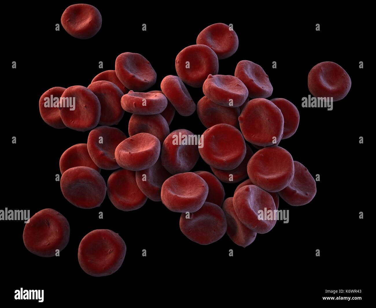 Topographical SEM (scanning Electron Microscope) close-up of oxygenated Red Blood Cells (Erythrocytes) piled up on dark grey surface background. Stock Photohttps://www.alamy.com/image-license-details/?v=1https://www.alamy.com/topographical-sem-scanning-electron-microscope-close-up-of-oxygenated-image159148195.html
RMK6WR43–Topographical SEM (scanning Electron Microscope) close-up of oxygenated Red Blood Cells (Erythrocytes) piled up on dark grey surface background.
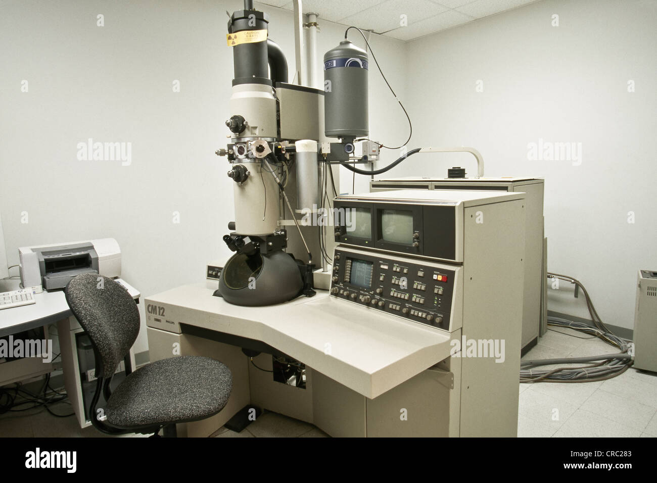 Philips CM12 Scanning/transmission Electron Microscope. Stock Photohttps://www.alamy.com/image-license-details/?v=1https://www.alamy.com/stock-photo-philips-cm12-scanningtransmission-electron-microscope-48823043.html
RMCRC283–Philips CM12 Scanning/transmission Electron Microscope.
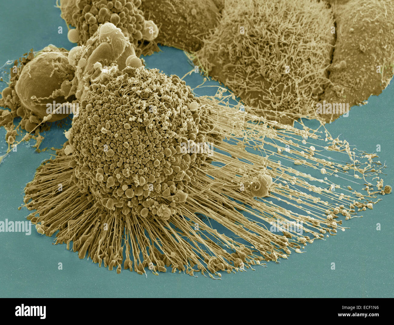 Scanning electron micrograph of an apoptotic HeLa cell. Zeiss Merlin HR-SEM. Stock Photohttps://www.alamy.com/image-license-details/?v=1https://www.alamy.com/stock-photo-scanning-electron-micrograph-of-an-apoptotic-hela-cell-zeiss-merlin-76548002.html
RFECF1N6–Scanning electron micrograph of an apoptotic HeLa cell. Zeiss Merlin HR-SEM.
 Iron oxide formations with sulphur and chlorine present, imaged with a scanning electron microscope Stock Photohttps://www.alamy.com/image-license-details/?v=1https://www.alamy.com/iron-oxide-formations-with-sulphur-and-chlorine-present-imaged-with-image69834381.html
RFE1H6D1–Iron oxide formations with sulphur and chlorine present, imaged with a scanning electron microscope
 Scanning electron microscope with EMF microanalysis Stock Photohttps://www.alamy.com/image-license-details/?v=1https://www.alamy.com/scanning-electron-microscope-with-emf-microanalysis-image591568486.html
RF2WAC7HX–Scanning electron microscope with EMF microanalysis
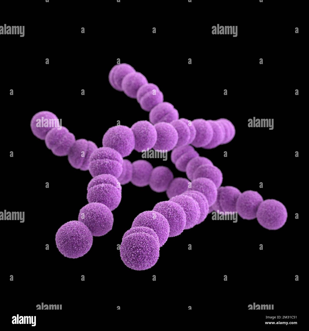 Group A streptococcus bacteria. STREP A Streptococcus pyogenes is a species of Gram-positive, aerotolerant bacteria in the genus Streptococcus. These bacteria are extracellular, and made up of non-motile and non-sporing cocci that tend to link in chains. This illustration depicted a 3D, computer-generated image, of a group of Gram-positive, Streptococcus pyogenes (group A Streptococcus) bacteria. The visualisation was based upon scanning electron microscopic (SEM) imagery. Optimised version of an image produced by the US Centers for Disease Control and Prevention / credit CDC /J.Oosthuizen Stock Photohttps://www.alamy.com/image-license-details/?v=1https://www.alamy.com/group-a-streptococcus-bacteria-strep-a-streptococcus-pyogenes-is-a-species-of-gram-positive-aerotolerant-bacteria-in-the-genus-streptococcus-these-bacteria-are-extracellular-and-made-up-of-non-motile-and-non-sporing-cocci-that-tend-to-link-in-chains-this-illustration-depicted-a-3d-computer-generated-image-of-a-group-of-gram-positive-streptococcus-pyogenes-group-a-streptococcus-bacteria-the-visualisation-was-based-upon-scanning-electron-microscopic-sem-imagery-optimised-version-of-an-image-produced-by-the-us-centers-for-disease-control-and-prevention-credit-cdc-joosthuizen-image500976141.html
RM2M31C51–Group A streptococcus bacteria. STREP A Streptococcus pyogenes is a species of Gram-positive, aerotolerant bacteria in the genus Streptococcus. These bacteria are extracellular, and made up of non-motile and non-sporing cocci that tend to link in chains. This illustration depicted a 3D, computer-generated image, of a group of Gram-positive, Streptococcus pyogenes (group A Streptococcus) bacteria. The visualisation was based upon scanning electron microscopic (SEM) imagery. Optimised version of an image produced by the US Centers for Disease Control and Prevention / credit CDC /J.Oosthuizen
 Unrecognizable scientist working with scanning electron microscope. Laboratory technician observing samples with a SEM. Stock Photohttps://www.alamy.com/image-license-details/?v=1https://www.alamy.com/unrecognizable-scientist-working-with-scanning-electron-microscope-laboratory-technician-observing-samples-with-a-sem-image428854030.html
RF2FWKYKA–Unrecognizable scientist working with scanning electron microscope. Laboratory technician observing samples with a SEM.
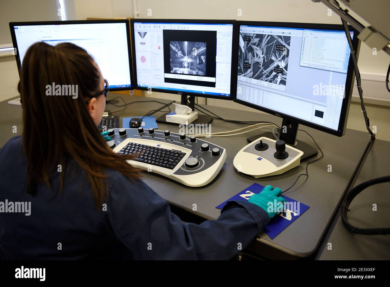 Zeiss EVO 15 Scanning Electron Microscope scanning station in a science lab. It is flexible variable pressure scanning electron microscope or SEM with Stock Photohttps://www.alamy.com/image-license-details/?v=1https://www.alamy.com/zeiss-evo-15-scanning-electron-microscope-scanning-station-in-a-science-lab-it-is-flexible-variable-pressure-scanning-electron-microscope-or-sem-with-image398273975.html
RM2E3XXEF–Zeiss EVO 15 Scanning Electron Microscope scanning station in a science lab. It is flexible variable pressure scanning electron microscope or SEM with
 Young female scientist in lab using scanning electron microscope Stock Photohttps://www.alamy.com/image-license-details/?v=1https://www.alamy.com/stock-photo-young-female-scientist-in-lab-using-scanning-electron-microscope-84830405.html
RFEX0A19–Young female scientist in lab using scanning electron microscope
 pressure valve Environmental Scanning Electron Microscope ESEM QuantaTM 250 FEG provides access to studies of wet biological samples, nano-bio compos Stock Photohttps://www.alamy.com/image-license-details/?v=1https://www.alamy.com/pressure-valve-environmental-scanning-electron-microscope-esem-quantatm-250-feg-provides-access-to-studies-of-wet-biological-samples-nano-bio-compos-image603831762.html
RM2X2AWG2–pressure valve Environmental Scanning Electron Microscope ESEM QuantaTM 250 FEG provides access to studies of wet biological samples, nano-bio compos
 Asbestos fibres under the electron scanning microscope Stock Photohttps://www.alamy.com/image-license-details/?v=1https://www.alamy.com/asbestos-fibres-under-the-electron-scanning-microscope-image331592797.html
RM2A7D9YW–Asbestos fibres under the electron scanning microscope
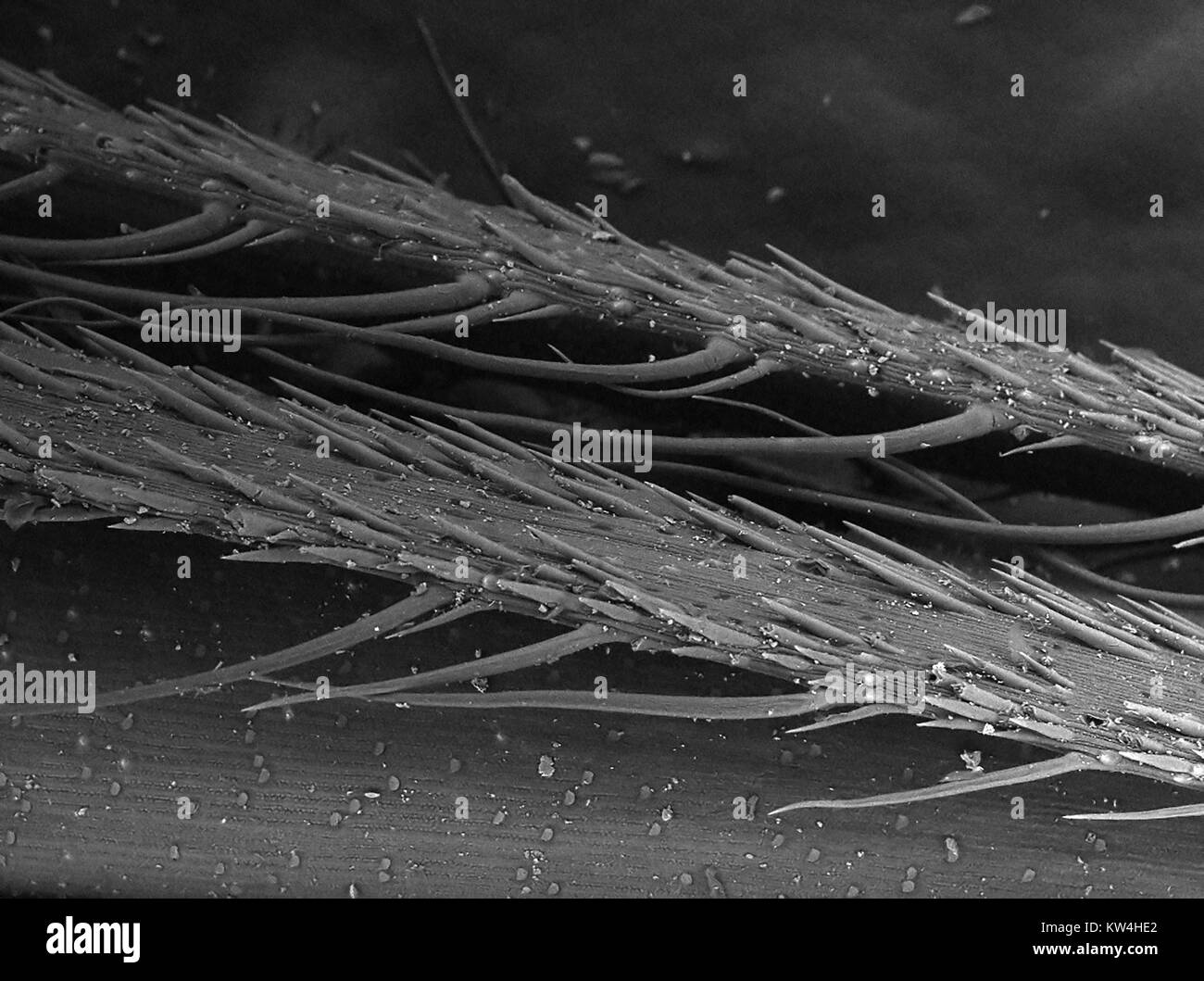 Scanning electron microscope (SEM) micrograph of the surface of a piece of foxtail grass (Hordeum murinum), showing microbarbs, at a magnification of 180x, 2016. Stock Photohttps://www.alamy.com/image-license-details/?v=1https://www.alamy.com/stock-photo-scanning-electron-microscope-sem-micrograph-of-the-surface-of-a-piece-170361242.html
RMKW4HE2–Scanning electron microscope (SEM) micrograph of the surface of a piece of foxtail grass (Hordeum murinum), showing microbarbs, at a magnification of 180x, 2016.
 Large Caterpillar: Gold coated and imaged in Scanning electron microscope Stock Photohttps://www.alamy.com/image-license-details/?v=1https://www.alamy.com/stock-photo-large-caterpillar-gold-coated-and-imaged-in-scanning-electron-microscope-71358916.html
RFE42K0M–Large Caterpillar: Gold coated and imaged in Scanning electron microscope
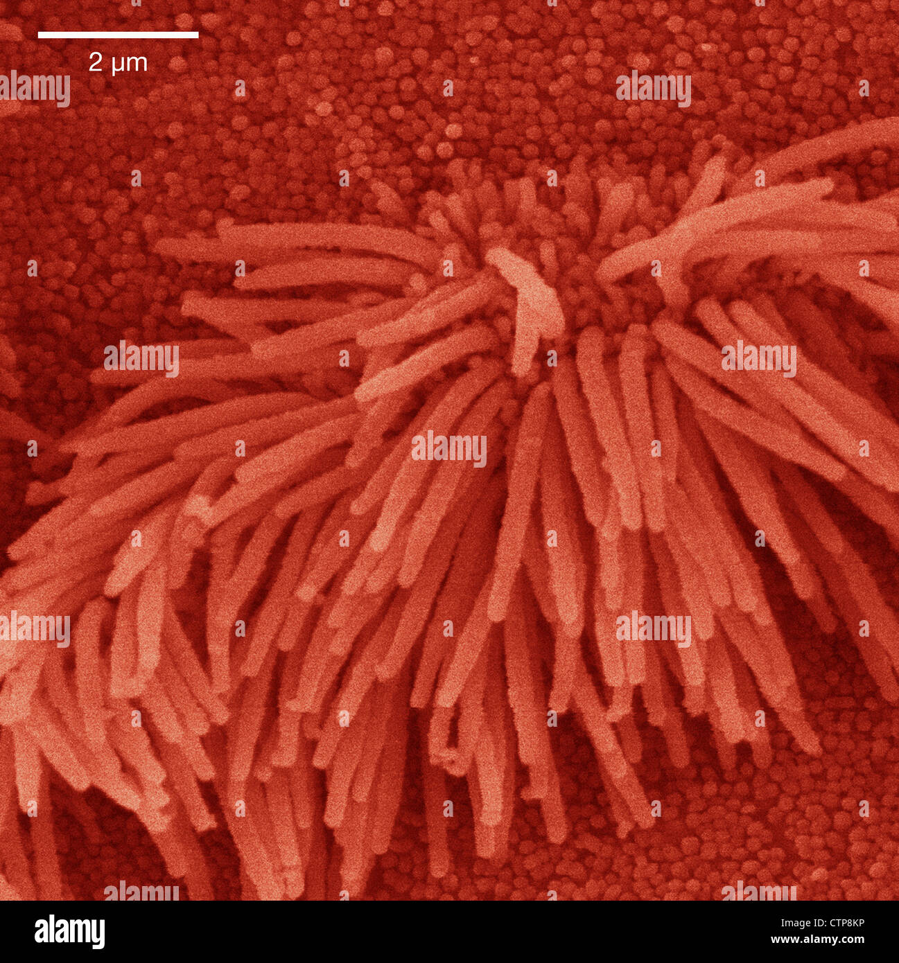 Scanning electron microscope image of lung trachea epithelium Stock Photohttps://www.alamy.com/image-license-details/?v=1https://www.alamy.com/stock-photo-scanning-electron-microscope-image-of-lung-trachea-epithelium-49662250.html
RMCTP8KP–Scanning electron microscope image of lung trachea epithelium
 Two scientists standing in analytical laboratory with scanning electron microscope Stock Photohttps://www.alamy.com/image-license-details/?v=1https://www.alamy.com/two-scientists-standing-in-analytical-laboratory-with-scanning-electron-image69861382.html
RFE1JCWA–Two scientists standing in analytical laboratory with scanning electron microscope
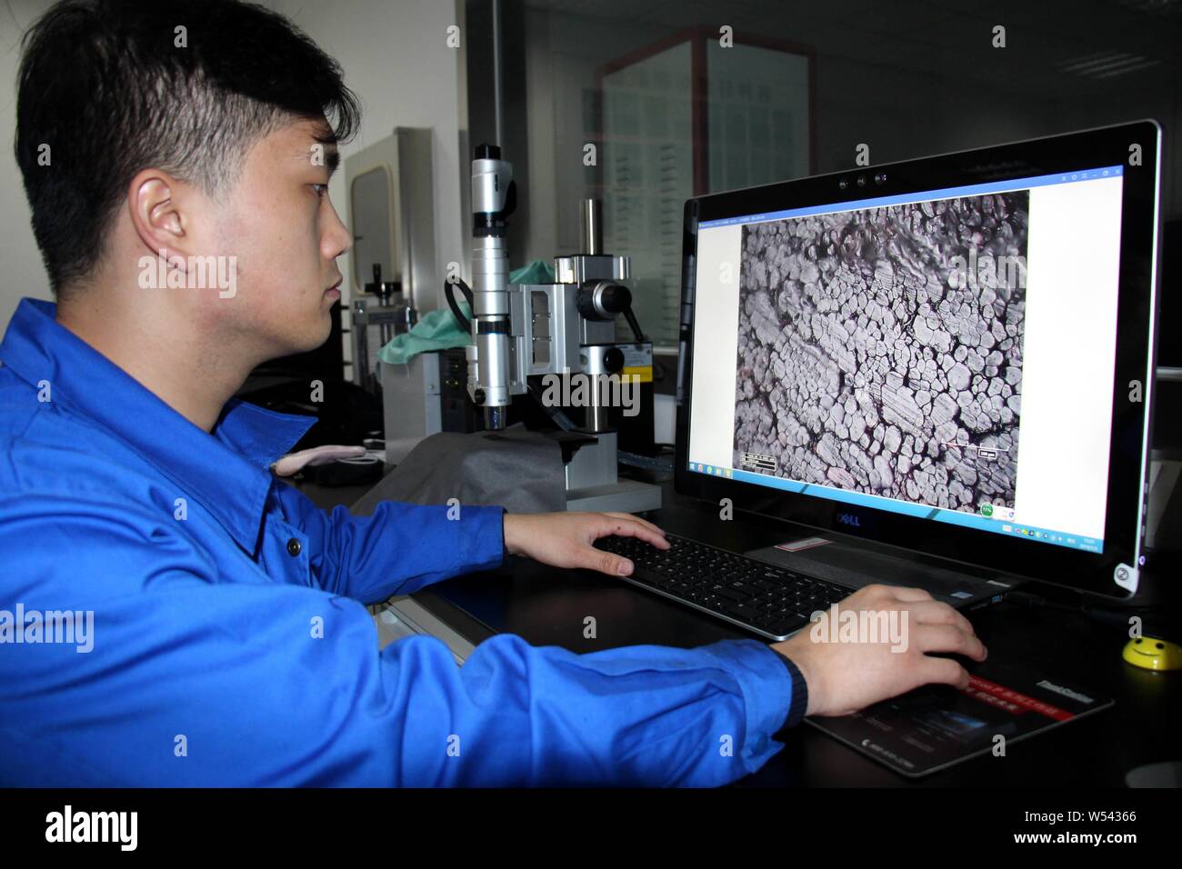 A Chines worker uses a scanning electron microscope to observe the structure of China's first far infrared conductive heating fiber developed based on Stock Photohttps://www.alamy.com/image-license-details/?v=1https://www.alamy.com/a-chines-worker-uses-a-scanning-electron-microscope-to-observe-the-structure-of-chinas-first-far-infrared-conductive-heating-fiber-developed-based-on-image261319134.html
RMW54366–A Chines worker uses a scanning electron microscope to observe the structure of China's first far infrared conductive heating fiber developed based on
 This scanning electron microscope image shows SARS-CoV-2 (round yellow particles) emerging from the surface of a cell cultured in the laboratory. SARS-CoV-2, also known as 2019-nCoV, is the virus that causes COVID-19. Image captured and colorized at Rocky Mountain Laboratories in Hamilton, Montana. Credit: NIAID Stock Photohttps://www.alamy.com/image-license-details/?v=1https://www.alamy.com/this-scanning-electron-microscope-image-shows-sars-cov-2-round-yellow-particles-emerging-from-the-surface-of-a-cell-cultured-in-the-laboratory-sars-cov-2-also-known-as-2019-ncov-is-the-virus-that-causes-covid-19-image-captured-and-colorized-at-rocky-mountain-laboratories-in-hamilton-montana-credit-niaid-image476706643.html
RM2JKFT57–This scanning electron microscope image shows SARS-CoV-2 (round yellow particles) emerging from the surface of a cell cultured in the laboratory. SARS-CoV-2, also known as 2019-nCoV, is the virus that causes COVID-19. Image captured and colorized at Rocky Mountain Laboratories in Hamilton, Montana. Credit: NIAID
 Surgical needle, SEM Stock Photohttps://www.alamy.com/image-license-details/?v=1https://www.alamy.com/surgical-needle-sem-image341959483.html
RF2AT9GPK–Surgical needle, SEM
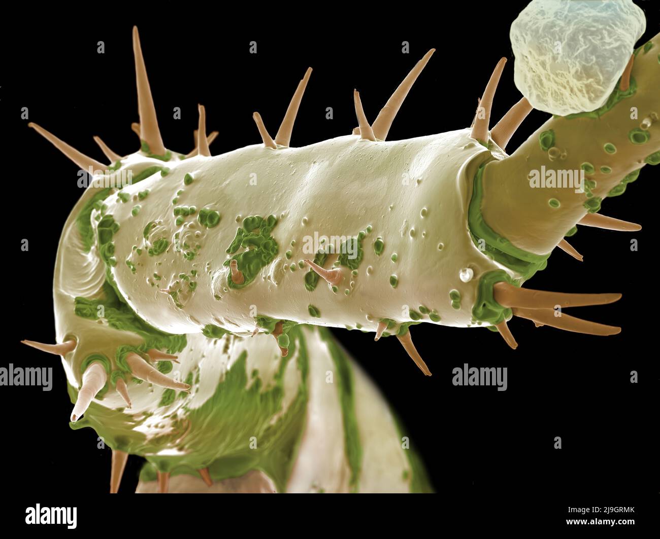 SEM Scanning Electron Microscope image of a Sandhopper, Sand Flea, amphipod Stock Photohttps://www.alamy.com/image-license-details/?v=1https://www.alamy.com/sem-scanning-electron-microscope-image-of-a-sandhopper-sand-flea-amphipod-image470581683.html
RM2J9GRMK–SEM Scanning Electron Microscope image of a Sandhopper, Sand Flea, amphipod
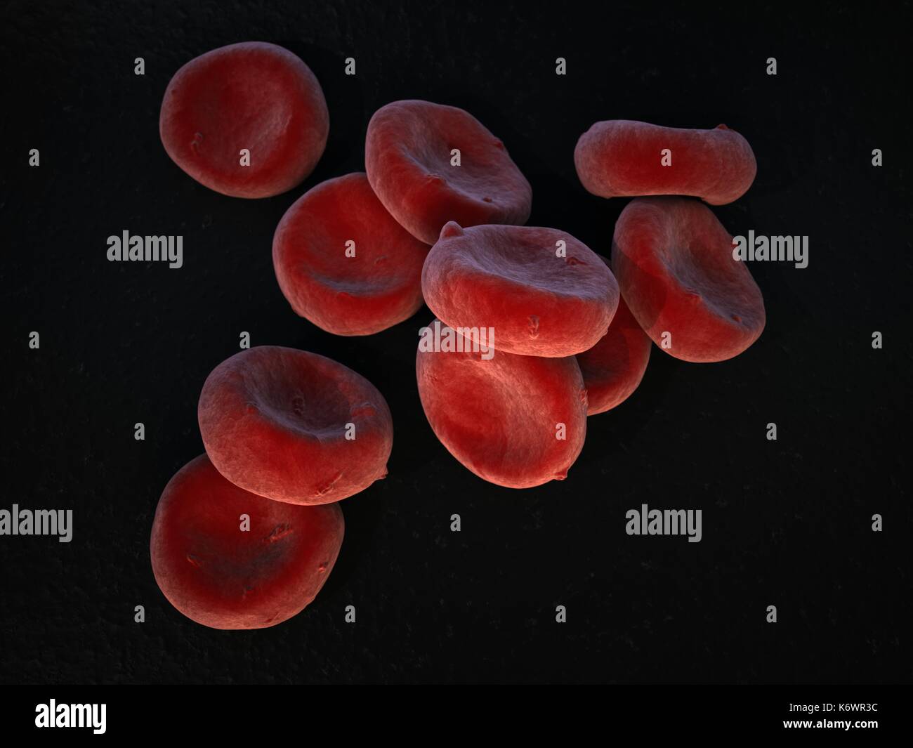 Extreme topographic close-up SEM (scanning Electron Microscope) of oxygenated Red Blood Cells (Erythrocytes) piled up on dark grey surface background. Stock Photohttps://www.alamy.com/image-license-details/?v=1https://www.alamy.com/extreme-topographic-close-up-sem-scanning-electron-microscope-of-oxygenated-image159148176.html
RMK6WR3C–Extreme topographic close-up SEM (scanning Electron Microscope) of oxygenated Red Blood Cells (Erythrocytes) piled up on dark grey surface background.
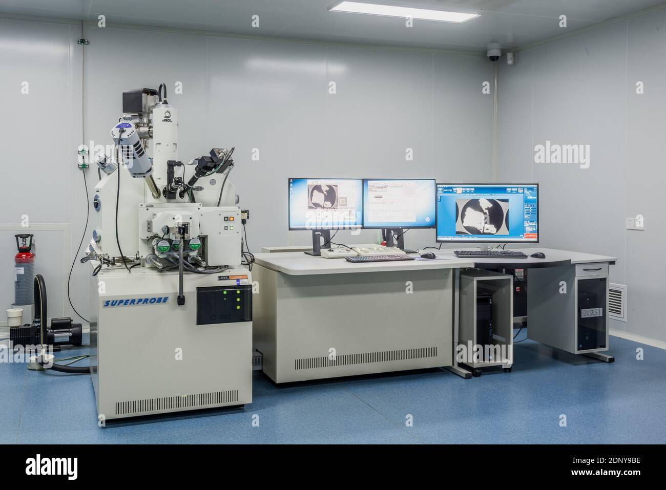 (201218) -- BEIJING, Dec. 18, 2020 (Xinhua) -- Photo taken on Nov. 27, 2020 shows the scanning electron microscope in a laboratory for moon samples at the National Astronomical Observatories of the Chinese Academy of Sciences in Beijing, capital of China. The moon samples collected by China's Chang'e-5 probe will be unsealed at the laboratory. (National Astronomical Observatories, CAS/Handout via Xinhua) Stock Photohttps://www.alamy.com/image-license-details/?v=1https://www.alamy.com/201218-beijing-dec-18-2020-xinhua-photo-taken-on-nov-27-2020-shows-the-scanning-electron-microscope-in-a-laboratory-for-moon-samples-at-the-national-astronomical-observatories-of-the-chinese-academy-of-sciences-in-beijing-capital-of-china-the-moon-samples-collected-by-chinas-change-5-probe-will-be-unsealed-at-the-laboratory-national-astronomical-observatories-cashandout-via-xinhua-image392135954.html
RM2DNY9BE–(201218) -- BEIJING, Dec. 18, 2020 (Xinhua) -- Photo taken on Nov. 27, 2020 shows the scanning electron microscope in a laboratory for moon samples at the National Astronomical Observatories of the Chinese Academy of Sciences in Beijing, capital of China. The moon samples collected by China's Chang'e-5 probe will be unsealed at the laboratory. (National Astronomical Observatories, CAS/Handout via Xinhua)
 A colorized scanning electron micrograph of a white blood cell eating an antibiotic resistant strain of Staphylococcus aureus ba Stock Photohttps://www.alamy.com/image-license-details/?v=1https://www.alamy.com/stock-photo-a-colorized-scanning-electron-micrograph-of-a-white-blood-cell-eating-76547752.html
RFECF1C8–A colorized scanning electron micrograph of a white blood cell eating an antibiotic resistant strain of Staphylococcus aureus ba
 Iron oxide formations with chlorine and sulphur present, imaged in a scanning electron microscope Stock Photohttps://www.alamy.com/image-license-details/?v=1https://www.alamy.com/iron-oxide-formations-with-chlorine-and-sulphur-present-imaged-in-image69834390.html
RFE1H6DA–Iron oxide formations with chlorine and sulphur present, imaged in a scanning electron microscope
 Mosquito front view taken with the scanning electron microscope Stock Photohttps://www.alamy.com/image-license-details/?v=1https://www.alamy.com/stock-photo-mosquito-front-view-taken-with-the-scanning-electron-microscope-14688568.html
RMAJBXJH–Mosquito front view taken with the scanning electron microscope
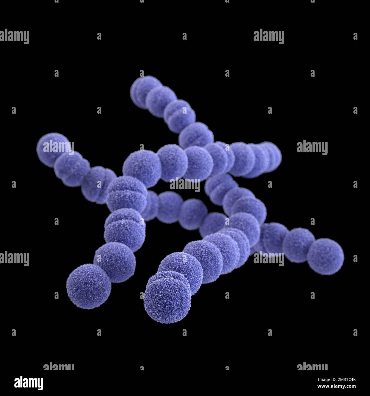 Group A streptococcus bacteria. STREP A Streptococcus pyogenes is a species of Gram-positive, aerotolerant bacteria in the genus Streptococcus. These bacteria are extracellular, and made up of non-motile and non-sporing cocci that tend to link in chains. This illustration depicted a 3D, computer-generated image, of a group of Gram-positive, Streptococcus pyogenes (group A Streptococcus) bacteria. The visualisation was based upon scanning electron microscopic (SEM) imagery. Optimised version of an image produced by the US Centers for Disease Control and Prevention / credit CDC /J.Oosthuizen Stock Photohttps://www.alamy.com/image-license-details/?v=1https://www.alamy.com/group-a-streptococcus-bacteria-strep-a-streptococcus-pyogenes-is-a-species-of-gram-positive-aerotolerant-bacteria-in-the-genus-streptococcus-these-bacteria-are-extracellular-and-made-up-of-non-motile-and-non-sporing-cocci-that-tend-to-link-in-chains-this-illustration-depicted-a-3d-computer-generated-image-of-a-group-of-gram-positive-streptococcus-pyogenes-group-a-streptococcus-bacteria-the-visualisation-was-based-upon-scanning-electron-microscopic-sem-imagery-optimised-version-of-an-image-produced-by-the-us-centers-for-disease-control-and-prevention-credit-cdc-joosthuizen-image500976131.html
RM2M31C4K–Group A streptococcus bacteria. STREP A Streptococcus pyogenes is a species of Gram-positive, aerotolerant bacteria in the genus Streptococcus. These bacteria are extracellular, and made up of non-motile and non-sporing cocci that tend to link in chains. This illustration depicted a 3D, computer-generated image, of a group of Gram-positive, Streptococcus pyogenes (group A Streptococcus) bacteria. The visualisation was based upon scanning electron microscopic (SEM) imagery. Optimised version of an image produced by the US Centers for Disease Control and Prevention / credit CDC /J.Oosthuizen
 Young man scientist working with scanning electron microscope. Laboratory technician observing samples with a SEM. Stock Photohttps://www.alamy.com/image-license-details/?v=1https://www.alamy.com/young-man-scientist-working-with-scanning-electron-microscope-laboratory-technician-observing-samples-with-a-sem-image427661779.html
RF2FRNJXY–Young man scientist working with scanning electron microscope. Laboratory technician observing samples with a SEM.
 Covid-19 under the microscope. Cartoon virus from scanning electron microscope. Spread of viruses by airborne droplets. Symbol of pandemic, lockdown, Stock Vectorhttps://www.alamy.com/image-license-details/?v=1https://www.alamy.com/covid-19-under-the-microscope-cartoon-virus-from-scanning-electron-microscope-spread-of-viruses-by-airborne-droplets-symbol-of-pandemic-lockdown-image466482193.html
RF2J2X2P9–Covid-19 under the microscope. Cartoon virus from scanning electron microscope. Spread of viruses by airborne droplets. Symbol of pandemic, lockdown,
 Scanning electron microscope SEM image of tiny wasp head. Stock Photohttps://www.alamy.com/image-license-details/?v=1https://www.alamy.com/stock-photo-scanning-electron-microscope-sem-image-of-tiny-wasp-head-14898121.html
RMAK668X–Scanning electron microscope SEM image of tiny wasp head.
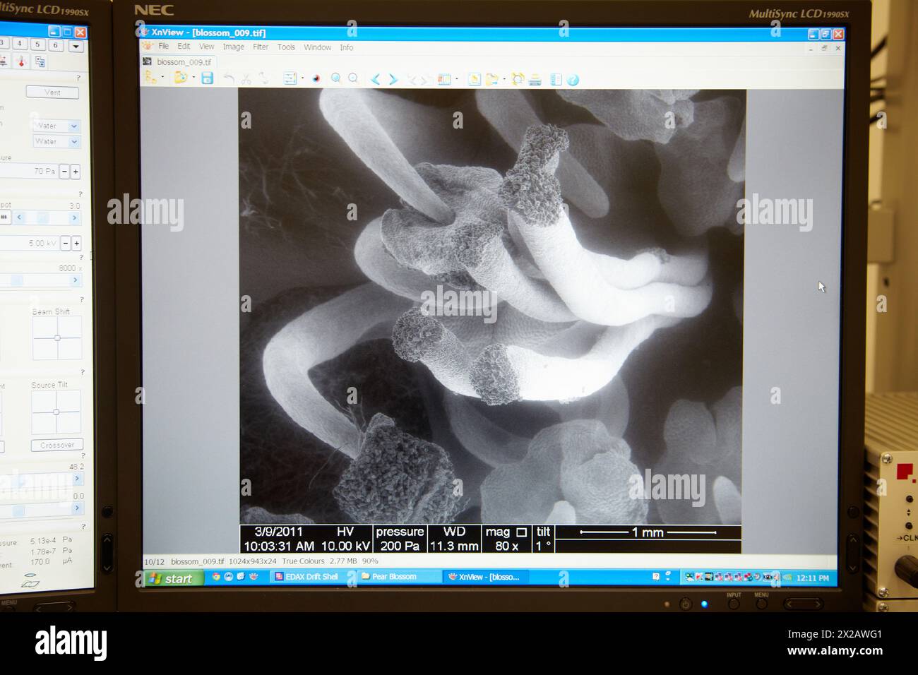 Pear blossom image, Environmental Scanning Electron Microscope ESEM QuantaTM 250 FEG provides access to studies of wet biological samples, nano-bio co Stock Photohttps://www.alamy.com/image-license-details/?v=1https://www.alamy.com/pear-blossom-image-environmental-scanning-electron-microscope-esem-quantatm-250-feg-provides-access-to-studies-of-wet-biological-samples-nano-bio-co-image603831761.html
RM2X2AWG1–Pear blossom image, Environmental Scanning Electron Microscope ESEM QuantaTM 250 FEG provides access to studies of wet biological samples, nano-bio co
 Virus in blood – among the red blood cells – Scanning Electron Microscopy stylized Stock Photohttps://www.alamy.com/image-license-details/?v=1https://www.alamy.com/stock-photo-virus-in-blood-among-the-red-blood-cells-scanning-electron-microscopy-47457092.html
RFCN5T04–Virus in blood – among the red blood cells – Scanning Electron Microscopy stylized
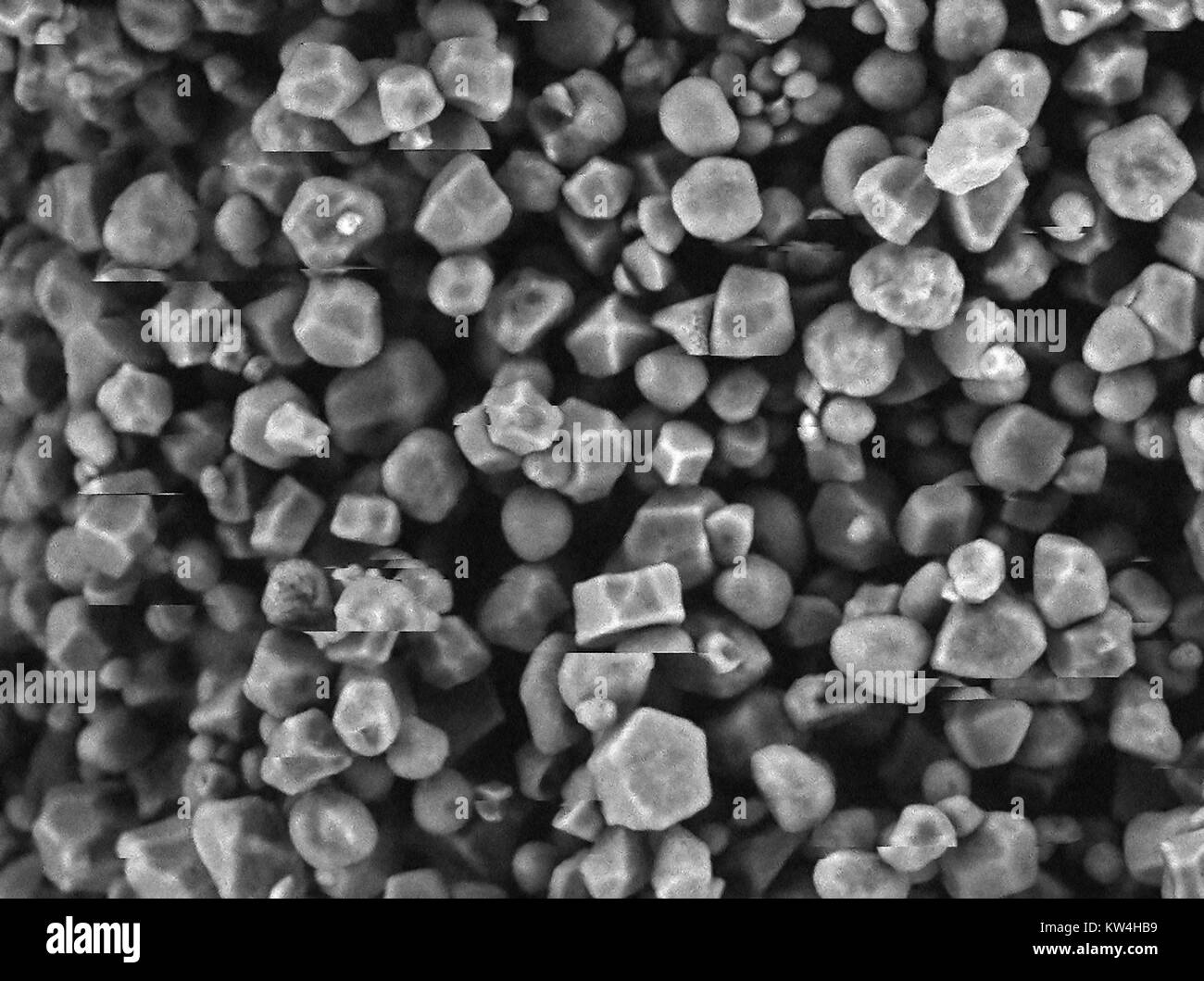 Scanning electron microscope (SEM) micrograph showing granules of corn starch, at a magnification of 1000x, 2016. Image shows artifacts related to charging effect on the sample. Stock Photohttps://www.alamy.com/image-license-details/?v=1https://www.alamy.com/stock-photo-scanning-electron-microscope-sem-micrograph-showing-granules-of-corn-170361165.html
RMKW4HB9–Scanning electron microscope (SEM) micrograph showing granules of corn starch, at a magnification of 1000x, 2016. Image shows artifacts related to charging effect on the sample.
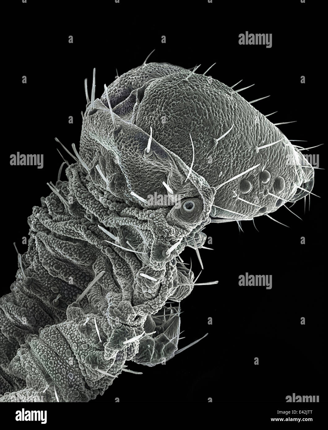 Large Caterpillar: Gold coated and imaged in Scanning electron microscope Stock Photohttps://www.alamy.com/image-license-details/?v=1https://www.alamy.com/stock-photo-large-caterpillar-gold-coated-and-imaged-in-scanning-electron-microscope-71358808.html
RFE42JTT–Large Caterpillar: Gold coated and imaged in Scanning electron microscope
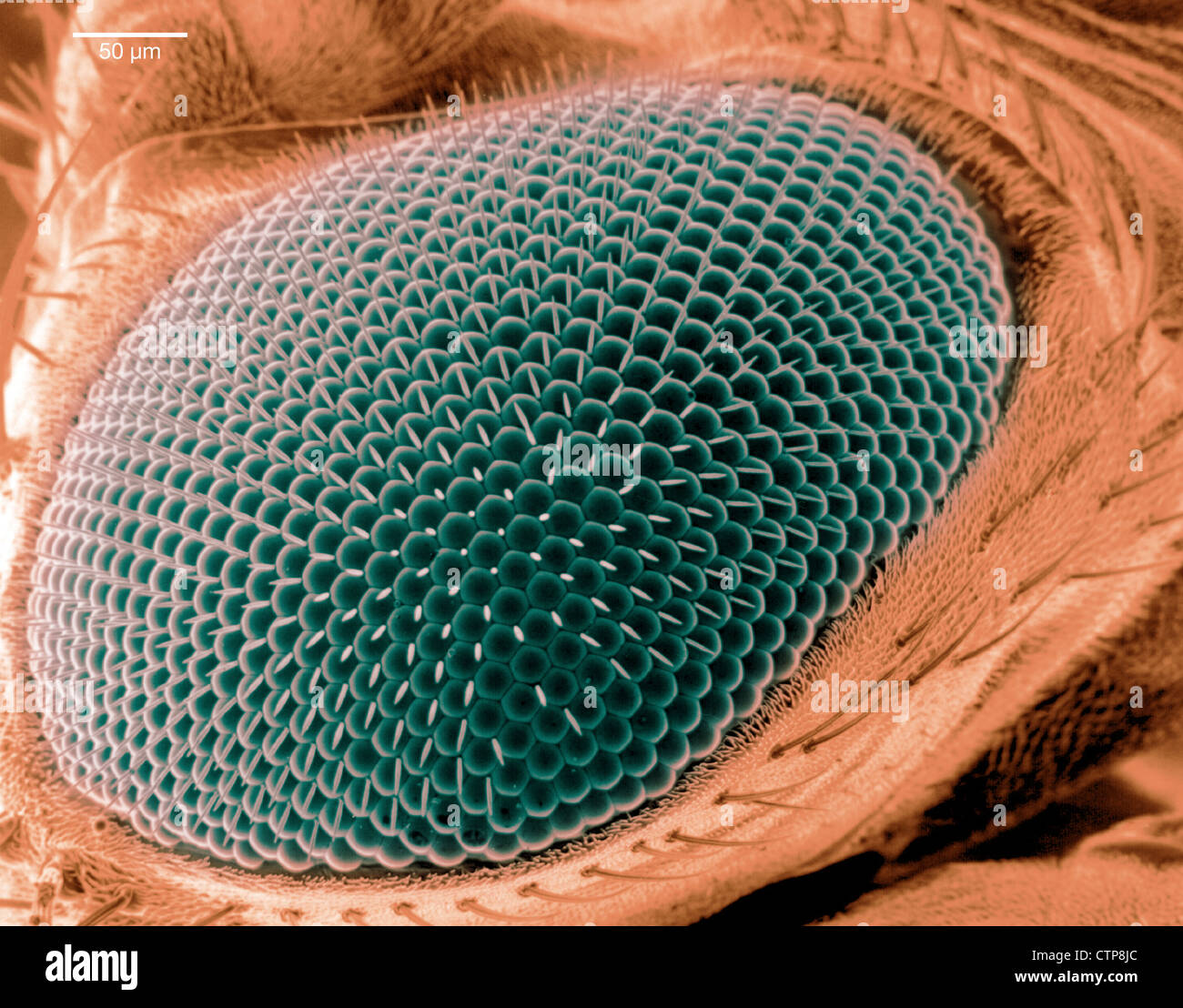 Scanning electron microscope image of an eye on a fruit fly. Stock Photohttps://www.alamy.com/image-license-details/?v=1https://www.alamy.com/stock-photo-scanning-electron-microscope-image-of-an-eye-on-a-fruit-fly-49662212.html
RMCTP8JC–Scanning electron microscope image of an eye on a fruit fly.
 Scientist standing in analytical laboratory with scanning electron microscope in foreground Stock Photohttps://www.alamy.com/image-license-details/?v=1https://www.alamy.com/scientist-standing-in-analytical-laboratory-with-scanning-electron-image69861387.html
RFE1JCWF–Scientist standing in analytical laboratory with scanning electron microscope in foreground
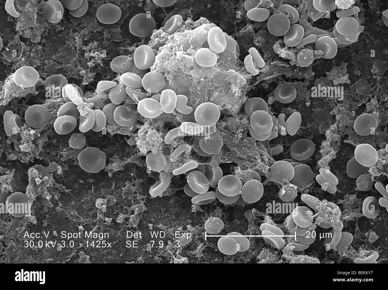 scanning electron microscope image of a blood clot showing red cells and fibrin coagulum Stock Photohttps://www.alamy.com/image-license-details/?v=1https://www.alamy.com/stock-photo-scanning-electron-microscope-image-of-a-blood-clot-showing-red-cells-17533355.html
RMB0EKY7–scanning electron microscope image of a blood clot showing red cells and fibrin coagulum
 This scanning electron microscope image shows SARS-CoV-2 (round yellow particles) emerging from the surface of a cell cultured in the laboratory. SARS-CoV-2, also known as 2019-nCoV, is the virus that causes COVID-19. Image captured and colorized at Rocky Mountain Laboratories in Hamilton, Montana. Credit: NIAID Stock Photohttps://www.alamy.com/image-license-details/?v=1https://www.alamy.com/this-scanning-electron-microscope-image-shows-sars-cov-2-round-yellow-particles-emerging-from-the-surface-of-a-cell-cultured-in-the-laboratory-sars-cov-2-also-known-as-2019-ncov-is-the-virus-that-causes-covid-19-image-captured-and-colorized-at-rocky-mountain-laboratories-in-hamilton-montana-credit-niaid-image476706632.html
RM2JKFT4T–This scanning electron microscope image shows SARS-CoV-2 (round yellow particles) emerging from the surface of a cell cultured in the laboratory. SARS-CoV-2, also known as 2019-nCoV, is the virus that causes COVID-19. Image captured and colorized at Rocky Mountain Laboratories in Hamilton, Montana. Credit: NIAID
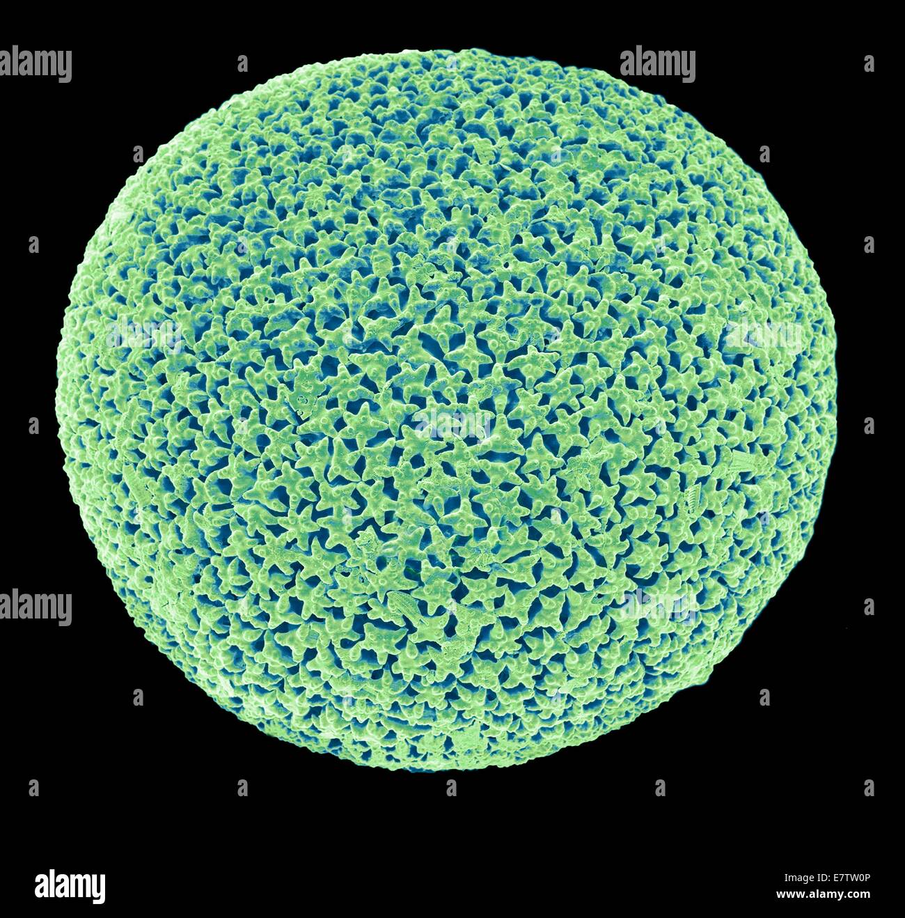 Orbulina. Coloured scanning electron micrograph (SEM) of the shell of the foraminiferan Orbulina sp. Foraminiferans are marine single-celled protists that construct and inhabit shells (tests), which are composed of several chambers. They are one of the ol Stock Photohttps://www.alamy.com/image-license-details/?v=1https://www.alamy.com/stock-photo-orbulina-coloured-scanning-electron-micrograph-sem-of-the-shell-of-73690534.html
RFE7TW0P–Orbulina. Coloured scanning electron micrograph (SEM) of the shell of the foraminiferan Orbulina sp. Foraminiferans are marine single-celled protists that construct and inhabit shells (tests), which are composed of several chambers. They are one of the ol
 SEM Scanning Electron Microscope image of a Sandhopper, Sand Flea, amphipod Stock Photohttps://www.alamy.com/image-license-details/?v=1https://www.alamy.com/sem-scanning-electron-microscope-image-of-a-sandhopper-sand-flea-amphipod-image470581234.html
RM2J9GR4J–SEM Scanning Electron Microscope image of a Sandhopper, Sand Flea, amphipod
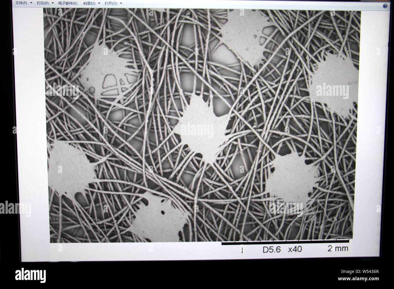 A Chines worker uses a scanning electron microscope to observe the structure of China's first far infrared conductive heating fiber developed based on Stock Photohttps://www.alamy.com/image-license-details/?v=1https://www.alamy.com/a-chines-worker-uses-a-scanning-electron-microscope-to-observe-the-structure-of-chinas-first-far-infrared-conductive-heating-fiber-developed-based-on-image261319151.html
RMW5436R–A Chines worker uses a scanning electron microscope to observe the structure of China's first far infrared conductive heating fiber developed based on
 Red Blood Cells (Erythrocytes) flowing in blood stream, SEM (scanning Electron Microscope) false rich red colored stylized depiction. Stock Photohttps://www.alamy.com/image-license-details/?v=1https://www.alamy.com/red-blood-cells-erythrocytes-flowing-in-blood-stream-sem-scanning-image159148213.html
RMK6WR4N–Red Blood Cells (Erythrocytes) flowing in blood stream, SEM (scanning Electron Microscope) false rich red colored stylized depiction.
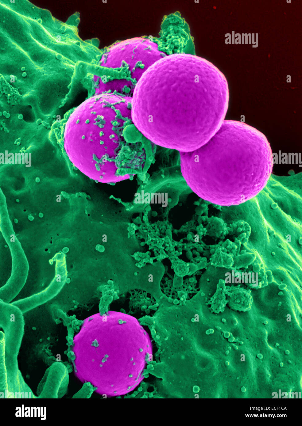 Scanning electron micrograph of a human neutrophil ingesting MRSA. Stock Photohttps://www.alamy.com/image-license-details/?v=1https://www.alamy.com/stock-photo-scanning-electron-micrograph-of-a-human-neutrophil-ingesting-mrsa-76547754.html
RFECF1CA–Scanning electron micrograph of a human neutrophil ingesting MRSA.
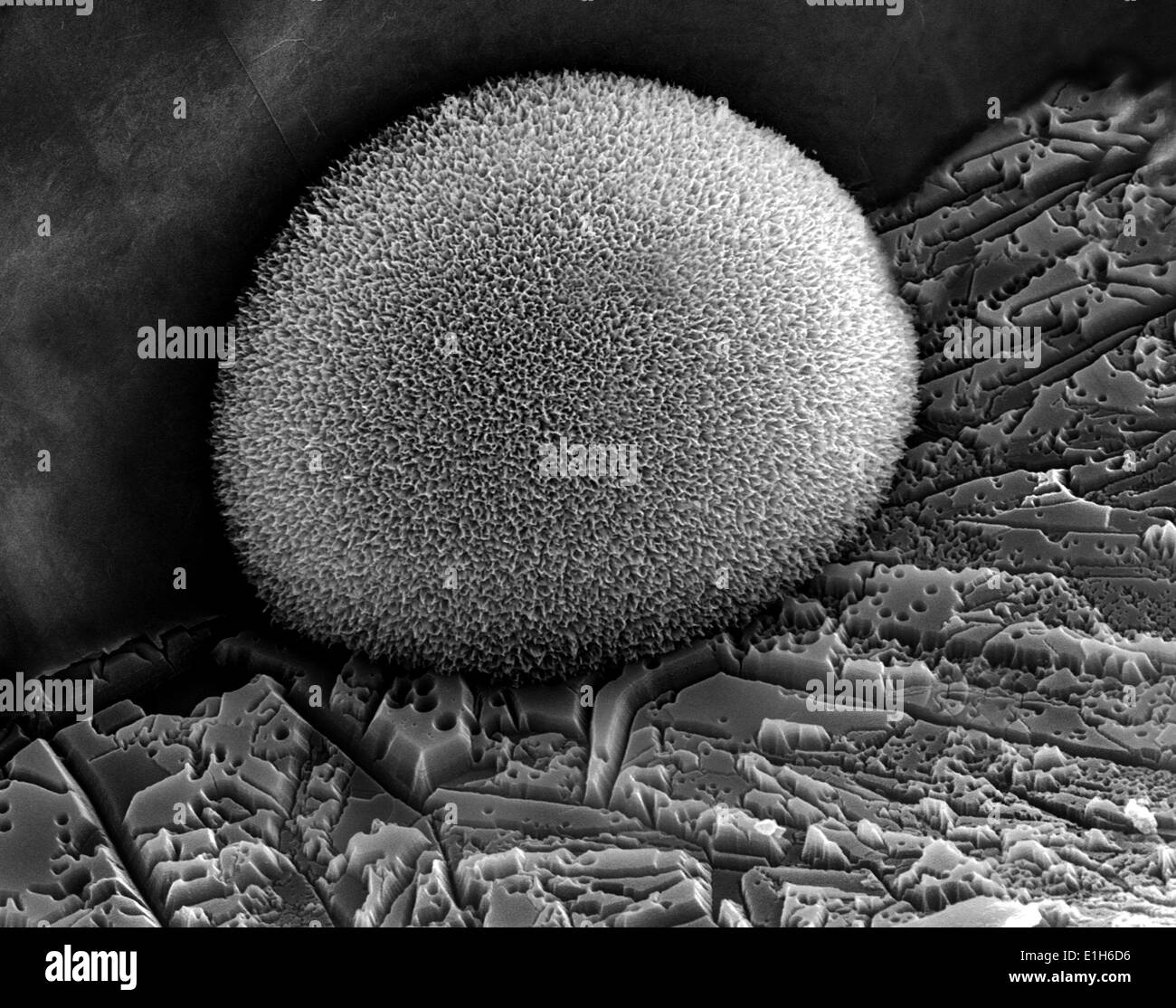 Iron oxide formations with sulphur and chlorine present, imaged in a scanning electron microscope Stock Photohttps://www.alamy.com/image-license-details/?v=1https://www.alamy.com/iron-oxide-formations-with-sulphur-and-chlorine-present-imaged-in-image69834386.html
RFE1H6D6–Iron oxide formations with sulphur and chlorine present, imaged in a scanning electron microscope
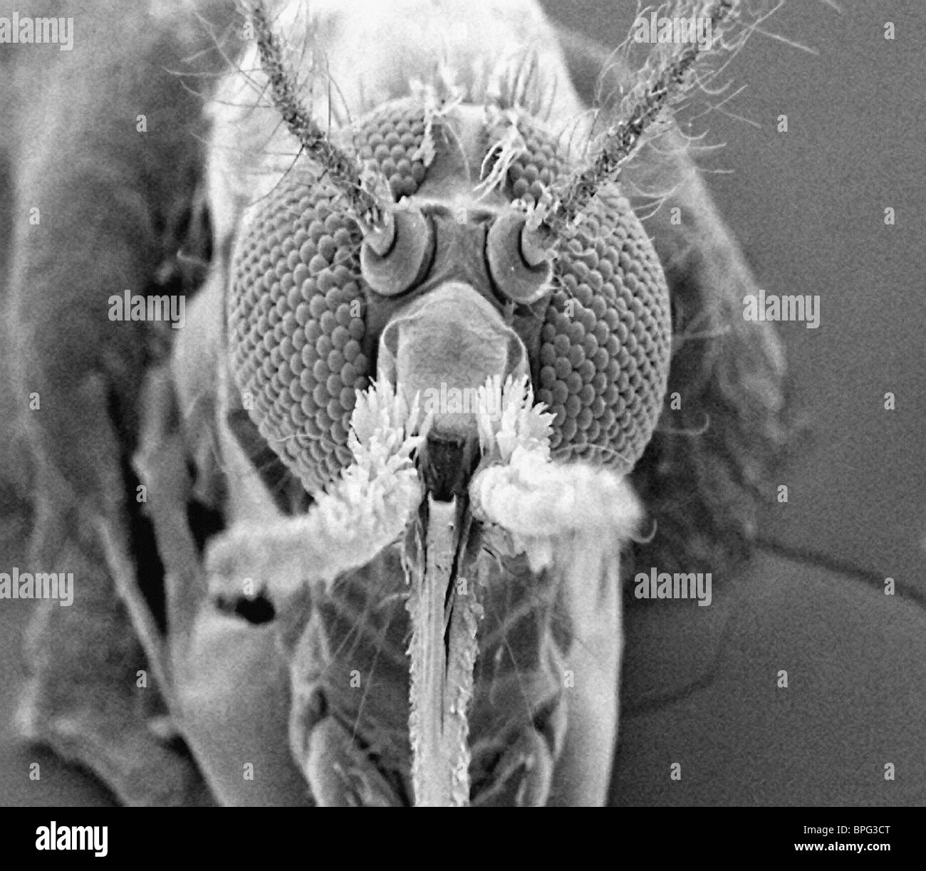 Mosquito - front view taken with the scanning electron microscope. Stock Photohttps://www.alamy.com/image-license-details/?v=1https://www.alamy.com/stock-photo-mosquito-front-view-taken-with-the-scanning-electron-microscope-31086744.html
RMBPG3CT–Mosquito - front view taken with the scanning electron microscope.