Medical Terminology Daily (MTD) is a blog sponsored by Clinical Anatomy Associates, Inc. as a service to the medical community. We post anatomical, medical or surgical terms, their meaning and usage, as well as biographical notes on anatomists, surgeons, and researchers through the ages. Be warned that some of the images used depict human anatomical specimens.

You are welcome to submit questions and suggestions using our "Contact Us" form. The information on this blog follows the terms on our "Privacy and Security Statement" and cannot be construed as medical guidance or instructions for treatment.


We have 391 guests and no members online


A Moment in History

Georg Eduard Von Rindfleisch

Georg Eduard Von Rindfleisch
(1836 – 1908)

German pathologist and histologist of Bavarian nobility ancestry. Rindfleisch studied medicine in Würzburg, Berlin, and Heidelberg, earning his MD in 1859 with the thesis “De Vasorum Genesi” (on the generation of vessels) under the tutelage of Rudolf Virchow (1821 - 1902). He then continued as a assistant to Virchow in a newly founded institute in Berlin. He then moved to Breslau in 1861 as an assistant to Rudolf Heidenhain (1834–1897), becoming a professor of pathological anatomy. In 1865 he became full professor in Bonn and in 1874 in Würzburg, where a new pathological institute was built according to his design (completed in 1878), where he worked until his retirement in 1906.

He was the first to describe the inflammatory background of multiple sclerosis in 1863, when he noted that demyelinated lesions have in their center small vessels that are surrounded by a leukocyte inflammatory infiltrate.

After extensive investigations, he suspected an infectious origin of tuberculosis - even before Robert Koch's detection of the tuberculosis bacillus in 1892. Rindfleisch 's special achievement is the description of the morphologically conspicuous macrophages in typhoid inflammation. His distinction between myocardial infarction and myocarditis in 1890 is also of lasting importance.

Associated eponyms

"Rindfleisch's folds": Usually a single semilunar fold of the serous surface of the pericardium around the origin of the aorta. Also known as the plica semilunaris aortæ.

"Rindfleisch's cells": Historical (and obsolete) name for eosinophilic leukocytes.

Personal note: G. Rindfleisch’s book “Traité D' Histologie Pathologique” 2nd edition (1873) is now part of my library. This book was translated from German to French by Dr. Frédéric Gross (1844-1927) , Associate Professor of the Medicine Faculty in Nancy, France. The book is dedicated to Dr. Theodore Billroth (1829-1894), an important surgeon whose pioneering work on subtotal gastrectomies paved the way for today’s robotic bariatric surgery. Dr. Miranda.

Sources:
1. "Stedmans Medical Eponyms" Forbis, P.; Bartolucci, SL; 1998 Williams and Wilkins
2. "Rindfleisch, Georg Eduard von (bayerischer Adel?)" Deutsche Biographie
3. "The pathology of multiple sclerosis and its evolution" Lassmann H. (1999)  Philos Trans R Soc Lond B Biol Sci. 354 (1390): 1635–40.
4. “Traité D' Histologie Pathologique” G.E.
Rindfleisch 2nd Ed (1873) Ballieres et Fils. Paris, Translated by F Gross


 "Clinical Anatomy Associates, Inc., and the contributors of "Medical Terminology Daily" wish to thank all individuals who donate their bodies and tissues for the advancement of education and research”.

Click here for more information


abebooks banner

bookplateink.com


Ascending aorta

Ascending aorta, anterior view
Ascending aorta

UPDATED: The ascending aorta is the first and most proximal portion of the aorta. About 5 cm. in length and 3 cm. in diameter at its origin, its proximal end begins at the superior aspect of the outflow tract of of the left ventricle, at the ventriculoaortic junction. 

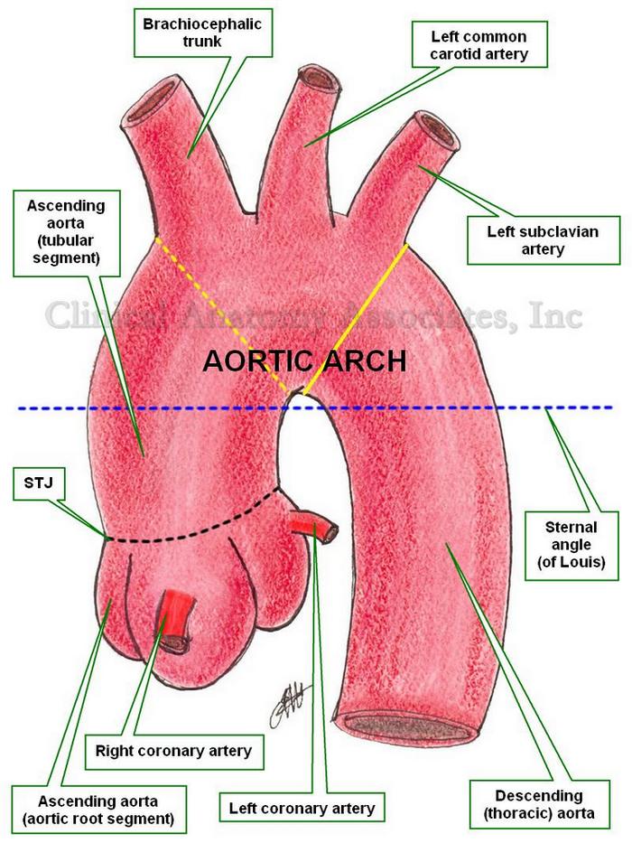
The ascending aorta ends superiorly at an imaginary horizontal plane (blue dotted line) that passes through the sternal angle (of Louis), continuing distally with the aortic arch. This is an important anatomical landmark, as many surgeons use as the superior border of the ascending aorta an oblique plane that passes proximal to the brachiocephalic trunk (yellow dotted line). Although this landmark could be useful in surgery, it is not anatomically correct.

Since the sternal angle (of Louis) also indicates the superior border of the pericardial sac, it can be said that the ascending aorta is completely intrapericardial, and in surgery the pericardial sac should be the anatomical landmark used to separate the ascending aorta from the aortic arch.

From its point of origin at the ventriculoaortic junction, the aorta presents with a dilated region where the aortic valve is located. The aortic valve is one of the two  semilunar valves of the heart, and the dilation of this region is caused by the presence of the sinuses of Valsalva. This dilated bulbous segment is known as the aortic root.

The dilated, sinus portion, or aortic root segment of the ascending aorta continues superiorly with the tubular portion of the ascending aorta. The area of transition between these two components is marked by a sharp crease known as the sinotubular junction (STJ). The dilation of the aortic root is caused by the presence of the sinuses of Valsalva, named after Antonio Maria Valsalva (1666 - 1723).

The tubular portion of the ascending aorta ascends with an inclination anteriorly and to the right. The ascending aorta presents with a slight anterior bulge causing the transverse section of this aortic segment to be slightly oval.

Only two arteries arise from the ascending aorta, both usually at the aortic root segment, just inferior to the STJ. These are the right coronary artery and the left coronary artery. There are anatomical variations where only one, or up to five different coronary arteries have been described.

At its origin, the aorta presents with semilunar folds of the serous surface of the pericardium. Also known as the ascending aortic folds, or Rindfleisch's folds, these are eponimically named after Georg Eduard Von Rindfleisch (1836 – 1908), a German pathologist and histologist of Bavarian nobility ancestry..

Image property of:CAA.Inc.Artist:Dr. E. Miranda