1. Name of the location of 90% of epistaxis
2. A genetic disorder that forms AV malformations in the skin, lungs, brain etc
3. Name of posterior vascular plexus in the nasal cavity causing posterior epistaxis
4. 1st line treatment for all epistaxis
5. The common brand name for anterior nasal packing
6. Chemical used in cautery sticks
7. Physically scaring complication of posterior nasal packing with foleys catheter
Coming soon..
Physiology of Hearing
Introduction
This page provides an overview of the physiology of human hearing to the level expected of students of medicine at Swansea University Medical School.
Hearing is the ability to perceive sound. It is achieved through the ear, which collects and amplifies vibrations in air before transducing these into neural activity that the brain can understand. This neural activity passes up, via the brain stem, to the cortex of the brain where it is processed and the sound is experienced. Sound collection, amplification, and transduction are discussed below.

The outer ear
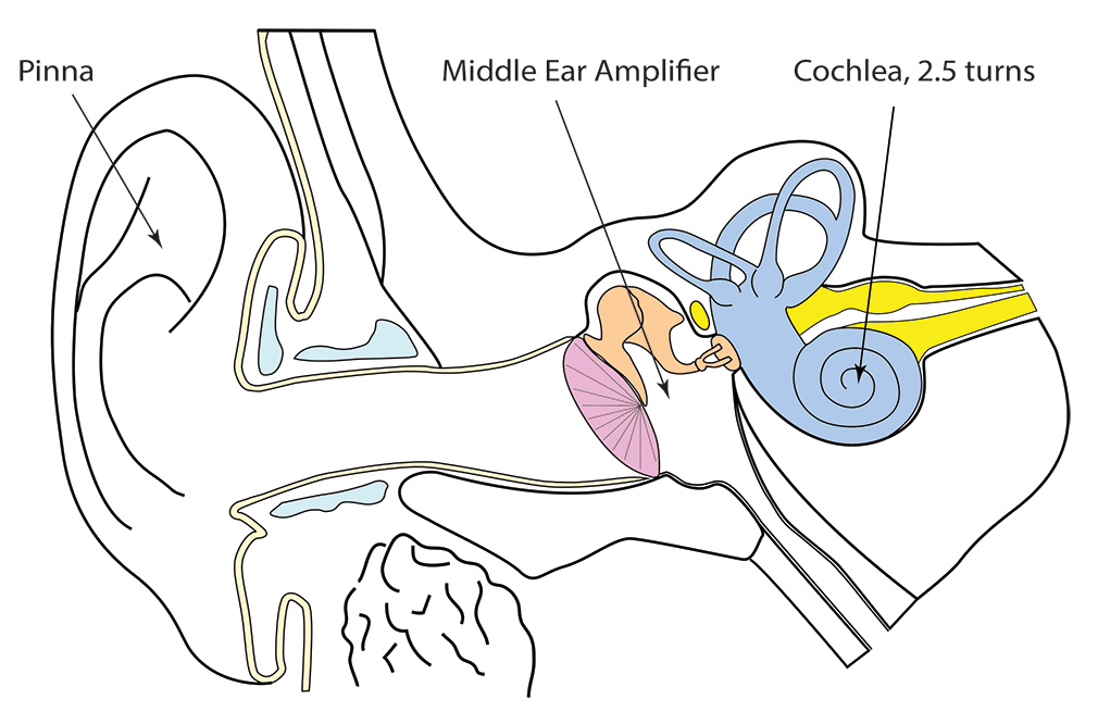
The shape of the outer ear has evolved to collect and direct sound towards the ear drum. The pinna and the external acoustic canal also amplify sounds and, because there is an ear on either side of the head, they allow us to detect the direction from which sound is coming.
The pinna, with its complex shape of folds and fossae is principally responsible for determining the source of sound when that sound is directly in front of, above or behind the head.
Detecting the source of sound from either side of the head is achieved through inter-aural intensity and latency differences. If sound is coming from your right side, as in the image below, it is slightly louder in the right ear than it is in the left ear. The head casts a sound shadow and the difference is perceptible. This works best for higher frequency sounds due to their short wavelength.
The source of sounds of lower frequency can still be detected, of course. In this case, it is the time of arrival of the sound that is used by the brain: sound from a source to the right of the head will reach the right ear before it gets to the left ear and, although the difference is very small, the brain is able to use this information to determine the direction from which the sound is coming.

The middle ear
The middle ear is an air-filled space containing three ossicles. These link the ear drum to the inner ear. It is the most medial bone, the stapes, which articulates with the oval window and vibrations of the stapes are transmitted directly to the perilymph in the inner ear through it.
The middle ear amplifies sound and directs it towards the fluid filled cochlea. Without it, 99.9% of the energy of sound would be lost. There are three mechanisms by which the transformer works but the most important is the area difference between the ear drum and the oval window. This difference amplifies sound 17-fold.
The levers mechanism of the ossicles of the middle ear has a very modest effect, but it adds to the gain produced by the ratio of drum to oval window area such that, overall, sound is amplified 20-fold. The third mechanism, the curved membrane buckling mechanism, is beyond the scope of this course.

Middle ear gain is frequency specific: 20dB between 250 and 500Hz; 27dB at 1kHz; 18dB at 2kHz and declining further with higher frequency.
Inner ear
The inner ear transduces the vibrations imparted to it by the stapes into neural activity. The process is called transduction and occurs in the Organ of Corti within the scala media. The cochlea also amplifies the response to sound when it is healthy.
The cochlear is a tubular structure consisting of three canals called scalae: s.vestibuli, s.media, and s. tympani. The scala vestibuli and tympani contain perilymph while the scala media contains endolymph. It is the scala media (sometimes called the cochlear duct) that contains the neuro-epithelium called the Organ of Corti.
The whole system is tapered and coiled into 2.5 turns so that the cochlea looks like a snail shell. A cross section through one of the turns of the cochlea is shown below.

The next diagram below shows the cochlea unravelled. The three scalae can be seen and it is evident that the scala vestibuli is continuous with the scala tympani. One becomes the other at a point called the helicotrema at the apex of the spiral system.
Sound enters the inner ear through the oval window via the stapes where it sets the perilymph (here in blue) vibrating. These vibrations are transmitted to the basilar membrane which vibrates in turn. Due to its physical properties, high-frequency sounds make the basilar membrane move most near the stapes (at the base of the cochlea) and low frequencies make it vibrate near its apex.

Just as in the vestibular system, the important basic unit of the Organ of Corti is the hair cell. These are lined up along the basilar membrane in rows, there being three rows of outer hair cells and one row of inner hair cells. Outer and inner refer to the centre of the cochlea. Each of the hair cells is topped by a tuft of stereocilia and these are embedded into an acellular, collagenous tectorial membrane.

Cross section through the Organ of Corti. The outer hair cells (pink) receive efferent input from the brainstem. They are involved in refining the tuning and sensitivity of the adjacent inner hair cells through their effect on the tectorial membrane. They act as a cochlear amplifier.
The single row of inner hair cells (dark purple) send afferent information to the brain stem.

The hair cells in the Organ of Corti have their support attached to the basilar membrane. The cilia at the apex of these cells are embedded into the tectorial membrane. When the basilar membrane moves up and down a shearing force is applied to the stereocilia because of the relative movement of their two attachments and this causes mechanically-gated membrane ion channels to open and allow potassium ions to move down their electrical gradient into the hair cell. In turn, calcium channels open and calcium enters the cell and this triggers exocytosis of neurotransmitter into the cleft between the cell and its neurone. The nerve fires.
Neural pathways
There are two pathways involved with hearing: the Classical Auditory Pathway (shown below) and the Non-Classical Pathway. In the Classical Pathway, cochlear nerve fibres run from the hair cell to the dorsal cochlear nucleus. From there, most fibres cross the midline to relay in the Superior Olive. Some fibres relay in the ipsilateral olive too. Both ipsilateral and contralateral Superior Olives convey impulses via the medial lemniscus to the inferior colliculus and then the medial geniculate body of the thalamus. From there, fibres pass via the auditory radiation into the Auditory Cortex.
The Non-Classical Auditory Pathway is far more diffuse and synapses with multiple other sensory inputs. It is not one that we study in Swansea.


