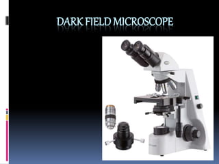
Dark field microscope
- 2. Dark-field microscopy produces an image with a dark background
- 3. How it works
- 4. DEFINATION Dark Field Microscopy is a technique used to observe unstained samples causing them to appear brightly lit against a dark, almost purely black, background.
- 5. A dark field microscopy is used to examine live microorganisms that either invisible in the ordinary light microscope, cannot be stained by standard methods, or are so distorted by staining that their characteristics then cannot be identified.
- 6. Contd… Instead of normal condenser, dark field microscope uses dark field condenser that contain a opaque disc.The disc blocks light that would enter the lens directly, only the light is reflected off the specimen enters the objective lens. Because there is no background light, the specimen appears light against black background- the dark field.
- 8. PRINCIPLE The dark ground microscope creates a contrast between object and surrounding field, such that, the background is dark and the object is bright The objective and the ocular lenses used in the dark ground microscope are same as that of ordinary light microscope.
- 9. Contd.. Special condenser is used, which prevents the transmitted light from directly illuminating the specimen Only oblique scattered light reaches the specimen and passes onto the lens system causing the object to appear bright against a dark background.
- 11. Uses of Dark field microscopy Diagnosis of Syphilis (Treponema pallidum). Viewing blood cells. Viewing bacteria. Viewing different types of algae. Viewing hairline metal fracture. Viewing diamonds and other precious stones. Viewing shrimp or other invertebrates
- 12. Advantages A dark field microscope is ideal for viewing unstained object, transparent and absorb little or no light. These specimens often have similar refractive indices as their surroundings, making them hard to distinguish with other illumination techniques.
- 13. Contd.. Dark field m/s used in research of live bacterium, as well as mounted cells and tissues. It is useful in examining external details, such as outlines, edges and surface defects than internal structures. Dark field is used study marine organisms such as algae ,plankton, diatoms, insects, as well as some minerals and crystals, thin polymers and some ceramics.
- 14. Contd.. Dark field has regained its popularity when combined with other illumination techniques, such as fluorescence, which widens its possible employment in certain fields. It is useful in examining external details, such as outlines, edges and surface defects than internal structure. .
- 15. Disadvantages, A specimen that is not thin enough or its density differs across the slide, may appear to have artefacts throughout the image. The preparation and quality of the slides can grossly affect the contrast and accuracy of a dark field image. You need to take special care that the slide, stage, nose and light source are free from small particles such as dust, as these will appear as part of the image.
- 16. Contd… You need to take special care that the slide, stage, nose and light source are free from dust, as these will appear as part of the image. Similarly, if you need to use oil or water on the condenser or slide, it is impossible to avoid air bubbles. These liquid bubbles will cause image degradation, and distortion .
- 17. Image of sugar crystals using Dark field microscope
- 21. Bacteria like structure in human blood
- 22. Submitted to, Dr Shilpa Dep’t of VPP Submitted by, Sahithya C.P Dep’t of VPT
- 23. Thank you


