This document discusses regurgitant murmurs, focusing on aortic regurgitation (AR) and mitral regurgitation (MR). It covers the etiology, pathophysiology, epidemiology, presentation, workup, management, and natural history of both conditions. For chronic MR, the left ventricle undergoes eccentric hypertrophy to compensate for the increased preload. Severe MR is defined by a regurgitant volume over 60mL/beat or an effective regurgitant orifice area over 0.40cm2. Surgery is generally recommended for symptomatic patients or those with progressive left ventricular dysfunction.
1) Pulmonary thromboembolism (PTE) is difficult to diagnose but can be life-threatening, with mortality rates around 30% in haemodynamically unstable patients.
2) PTE is a common cardiovascular emergency caused by emboli blocking pulmonary arteries, and has high morbidity and mortality rates.
3) Autopsy studies have found PTE to be the actual cause of death in up to 15% of hospitalized patients and the leading cause of unexpected in-hospital deaths.
AR, or aortic regurgitation, occurs when the aortic valve does not close properly, allowing blood to flow backward into the left ventricle. It can be caused by damage to the aortic valve leaflets or distortion/dilation of the aortic root. In developed countries, the most common causes are aortic root dilation or a congenital bicuspid aortic valve. AR is more common in men than women. Symptomatic patients or asymptomatic patients with reduced ejection fraction or increased left ventricular dimensions require surgical treatment such as aortic valve replacement. The prognosis depends on symptoms and left ventricular function, with asymptomatic patients having normal ejection fraction having an excellent long-term prognosis.
Aortic regurgitation occurs when the aortic valve does not close properly, allowing blood to flow back into the left ventricle. It can be caused by conditions that damage the aortic valve such as rheumatic fever or a congenital heart defect. Symptoms may include breathlessness, fatigue, and chest pain. Diagnosis is made through echocardiogram which can assess the severity. Treatment depends on severity but may involve lifestyle changes, medications, or aortic valve replacement surgery if symptoms worsen or damage to the heart progresses. Prognosis depends on severity and treatment, with severe untreated cases having a high risk of heart failure or sudden cardiac death.
comprehensive presentation on 2D echo use in ICu set up. helpful in finding causes of shock and also in monitoring of fluid status in critically ill patients.
Constrictive pericarditis is caused by scarring and thickening of the pericardium, restricting cardiac filling. It is diagnosed using echocardiography which shows septal bounce, exaggerated mitral inflow, and hepatic vein reversal. While similar to restrictive cardiomyopathy, constrictive pericarditis shows increased ventricular interaction and respiratory effects on cardiac physiology. Surgical pericardiectomy is usually required for treatment but is high risk, with post-op complications common. Long-term survival depends on the underlying cause of constriction.
1. Acyanotic congenital heart defects like ventricular septal defects (VSD), atrial septal defects (ASD), and atrioventricular septal defects involve left-to-right shunting that can cause pulmonary overcirculation and congestion over time.
2. During anesthesia for repairs of these defects, careful attention must be paid to balancing systemic and pulmonary vascular resistances to avoid increasing or decreasing the shunt. Inhalational induction is relatively contraindicated due to risk of shunt reversal and cyanosis.
3. Intravenous induction with ketamine or etomidate is preferred for acyanotic left-to-right shunt defects to gradually control hemodynamics
1. Mitral stenosis is most commonly caused by rheumatic fever and results in thickening and calcification of the mitral valve, reducing the valve orifice area and obstructing blood flow from the left atrium to ventricle.
2. The pathophysiology involves elevated left atrial pressure, pulmonary hypertension, and reduced cardiac output. Symptoms range from easy fatigability to pulmonary edema.
3. Physical exam findings include an opening snap, rumbling diastolic murmur, and signs of right heart failure in severe cases. Severity is graded based on orifice area, pulmonary artery pressure, and NYHA functional
This document provides information on congestive cardiac failure (CCF), including its definition, pathophysiology, clinical features, investigations, and management. CCF occurs when the heart muscle is weakened and cannot maintain adequate cardiac output. The pathophysiology involves changes in preload, afterload, and contractility that decrease cardiac output. Compensatory mechanisms initially help but later worsen symptoms. Clinically, CCF presents with dyspnea, edema, elevated JVP, hepatomegaly, and other signs. Investigations include BNP, ECG, echocardiogram. Management focuses on treating the underlying cause, reducing preload/afterload, and improving contractility. Diuretics, ACE inhibitors, beta
Peri op management of mitral stenosis patient coming for non cardiac surgeryanaesthesiaESICMCH
Mitral stenosis is a chronic mechanical obstruction of the left ventricular inflow caused by narrowing of the mitral valve orifice. This document discusses the anesthetic considerations for a patient with mitral stenosis undergoing non-cardiac surgery. It covers the pathophysiology of mitral stenosis, preoperative evaluation and optimization of the patient, and intraoperative anesthetic goals of maintaining normal hemodynamics while avoiding tachycardia, changes in preload or afterload, and worsening of pulmonary hypertension.
This document discusses hypertensive heart disease and its effects on the heart. It notes that hypertensive heart disease can cause left ventricular hypertrophy, left ventricular dysfunction (both diastolic and systolic), heart failure, arrhythmias, conduction abnormalities, coronary heart disease, and aortic regurgitation. It provides details on left ventricular hypertrophy including its classification, regression, diagnosis, and risks. It also discusses how hypertension can lead to left ventricular diastolic and systolic dysfunction as well as heart failure. The document outlines treatment options for regressing left ventricular hypertrophy, treating diastolic dysfunction and heart failure, and treating left ventricular systolic dysfunction.
Asymptomatic Severe Aortic Stenosis – Cardiologist’s Confusion and Surgeon’s ...Imran Ahmed
1) The management of asymptomatic severe aortic stenosis is complex due to uncertainties in assessing risk.
2) Exercise stress testing and biomarkers like BNP can help identify higher-risk asymptomatic patients who may benefit from earlier surgery.
3) Dobutamine stress echocardiography can help determine if low-flow, low-gradient aortic stenosis is true severe stenosis amenable to surgery or pseudostenosis better managed medically. Integrating all clinical and test results is important for individualizing treatment decisions in these patients.
1) The document discusses the use of echocardiography in evaluating patients in shock.
2) Basic echocardiography can readily identify the type of shock and guide diagnosis and management in most cases.
3) Key echocardiographic assessments include evaluating left ventricular size and function, identifying valvular pathology, assessing fluid status and volume responsiveness, and detecting causes of distributive, cardiogenic, obstructive, or hypovolemic shock.
4) Echocardiography is a useful first-line tool that can distinguish between different types of shock and serially monitor patients, helping clinicians optimize management of critically ill patients.
The document discusses guidelines for assessing diastolic dysfunction according to the ASE/EACVI 2016 guidelines. It defines diastolic dysfunction and describes the stages from grade I to grade IV. For each grade, it discusses the pathophysiology and key echocardiographic findings including mitral inflow patterns, tissue Doppler measurements, pulmonary vein flow, and left atrial size. The guidelines simplify the assessment of diastolic function into four grades based on parameters of left ventricular relaxation, left atrial pressure, mitral E/A ratio, E/e' ratio, pulmonary vein flow, and left atrial size.
This document discusses congenital heart disease in adults. It notes that 1 million adults in the US have congenital heart disease, with 20,000 more reaching adulthood each year due to increased survival of children with CHD. Common adult presentations of CHD include effort dyspnea, atrial fibrillation, and right heart failure. The document reviews the pathophysiology, clinical features, diagnostic evaluation, and management of various CHD lesions that may present in adulthood, such as atrial septal defects, ventricular septal defects, patent ductus arteriosus, tetralogy of Fallot, Ebstein's anomaly, and coarctation of the aorta. Surgical and percutaneous interventions are discussed
This document discusses valvular heart disease, focusing on aortic stenosis, mitral stenosis, aortic regurgitation, and mitral regurgitation. It covers the etiology, pathophysiology, presentation, evaluation, and management of each condition. For aortic and mitral stenosis, the goals are to recognize symptoms of severe disease and identify patients who need valve replacement. For regurgitant lesions, treatment focuses on supporting cardiac function medically until symptoms appear or structural deterioration occurs, then referring for surgical intervention. Echocardiography plays a key role in diagnosing severity and monitoring progression.
This document discusses pulmonary embolism (PE), including its risk factors, pathophysiology, diagnosis, and management. It notes that PE is a leading cause of cardiovascular death and hospital mortality. Rudolf Virchow identified three main factors that contribute to venous thrombosis and PE: venous stasis, hypercoagulability of blood, and endothelial injury. The pathophysiology of PE involves increased pulmonary vascular resistance and right heart strain or failure. Diagnosis involves assessing clinical risk factors, biomarkers like troponin and BNP, imaging tests like CTPA, and echocardiography. PE is classified as massive, submassive, or low-risk depending on the presence of hypotension and right heart dysfunction. Management involves he
This document discusses the pathogenesis and diagnosis of acute decompensated heart failure (ADHF). It defines ADHF and describes its epidemiology, including the high rates of hospitalization. Common comorbidities are hypertension, coronary artery disease, diabetes, and COPD. ADHF can be classified based on history, blood pressure, signs/symptoms, and ejection fraction. Causes include nonadherence, infection, ischemia, and arrhythmias. Pathophysiology involves impaired function, renal dysfunction, neurohormonal activation, and fluid overload leading to congestion. Evaluation includes symptoms, vital signs, jugular vein pressure, lung sounds, and edema. Labs include BNP/NT-proBNP, troponin,
The document discusses constrictive pericarditis, providing details on:
1) The pathology of constrictive pericarditis which involves thickening and scarring of the pericardium leading to loss of elasticity.
2) The pathophysiology of constrictive pericarditis where the inelastic pericardium constrains cardiac filling and prevents adaptation to volume changes.
3) Key diagnostic features of constrictive pericarditis seen on echocardiogram include septal bounce, rapid early diastolic mitral inflow, and increased mitral annular velocities that rise with inspiration.
This document provides an overview of cardiac ultrasound views and techniques. It discusses the four standard cardiac views: parasternal long axis (PLAX), parasternal short axis (SAX), apical four-chamber (A4C), and subcostal. For each view, it describes the probe placement, normal anatomy, and features of a good image. Common pathologies that can be identified include reduced contractility, pericardial effusion, pulmonary embolism, and regional wall motion abnormalities. Assessment of ejection fraction and identifying areas of abnormal wall motion are important for evaluating cardiac function and ischemia.
1. Congenital cyanotic heart disease refers to heart defects present at birth that result in low oxygen levels in the blood. They are classified as those with increased or decreased blood flow to the lungs.
2. Common types with increased pulmonary blood flow include persistent truncus arteriosus and transposition of the great arteries (TGA). Common types with decreased pulmonary blood flow include tricuspid atresia and pulmonary atresia with intact ventricular septum.
3. TGA accounts for 5-7% of congenital heart defects. It involves the aorta originating from the right ventricle and the pulmonary artery originating from the left ventricle. It requires mixing of blood between the pulmonary and systemic
A short update on aortic regurgitation drmohitmathur
Aortic regurgitation can be caused by primary valve disease or primary aortic root disease. In the initial stages, increased blood flow to the left ventricle maintains cardiac output but causes increased pressure over time. Eventually, the left ventricle's function deteriorates as blood flows backward, leading to further enlargement and heart failure if untreated. Surgical replacement of the aortic valve is often needed to treat severe chronic aortic regurgitation before left ventricular dysfunction progresses. Homoeopathic treatment may help prevent immune-mediated valve damage in some cases.
This document provides information on various cyanotic heart lesions including Tetralogy of Fallot, Transposition of the Great Arteries, Tricuspid Atresia, Ebstein's Anomaly and Total Anomalous Pulmonary Venous Return. It describes the anatomy, clinical features, diagnosis and management of these conditions. Key cyanotic heart lesions are characterized by mixing of oxygenated and deoxygenated blood resulting in central cyanosis.
This document discusses acyanotic congenital heart disease, which includes conditions with left-to-right shunts such as atrial septal defects (ASD), ventricular septal defects (VSD), and patent ductus arteriosus (PDA). It provides details on the pathophysiology, clinical presentation, investigations, and treatment options for each condition. ASDs can cause volume overloading of the right heart and pulmonary hypertension if large. VSDs similarly cause left ventricular volume overloading. PDAs shunt blood from the aorta to the pulmonary artery. All conditions are typically addressed through either surgical or catheter-based closure.
Tetralogy of Fallot (TOF) is the most common cyanotic congenital heart disease, occurring in approximately 1 in 3,000 live births. It involves four abnormalities - a ventricular septal defect, right ventricular outflow tract obstruction, right ventricular hypertrophy, and overriding of the aorta. Without surgical repair, only 10% of patients survive beyond 20 years of age. Clinical manifestations include cyanosis, clubbing of the fingers and toes, and hypoxic spells in infants. Diagnosis is made through echocardiography, chest x-ray, and cardiac catheterization. Treatment involves palliative shunt procedures for infants or complete repair surgery to close the ventricular septal defect and widen the right ventricular
This document discusses aortic stenosis, including its causes, symptoms, diagnostic studies, prognosis, and treatment options. It begins with an overview and definitions, then describes the main types and risk factors for aortic stenosis. Diagnostic tests like echocardiography are outlined for evaluating the severity of stenosis. The prognosis without treatment is explained. Treatment involves initially managing symptoms, with surgical aortic valve replacement as the definitive treatment for severe symptomatic stenosis. A case study is presented of an elderly woman found to have incidental moderate aortic stenosis while being evaluated for anemia.
This document summarizes HIV-associated neurological complications. It discusses several conditions that can affect the spinal cord, peripheral nervous system, and muscles in patients with HIV/AIDS, including:
1. Vacuolar myelopathy, a rare condition since ART that causes slowly progressive weakness.
2. Distal sensory polyneuropathy (DSP), the most common neuropathy affecting 30-50% of HIV patients, presenting as numbness and tingling.
3. Inflammatory demyelinating polyradiculoneuropathies like AIDP and CIDP, which develop during early infection and present with weakness.
4. Other rare conditions like lumbosacral polyradiculo
1) Acute rheumatic fever is an autoimmune disease that can develop as a complication of untreated strep throat. It mainly affects children aged 5-14 and can cause inflammation of the heart, joints, brain, and skin.
2) The presentation and diagnosis is based on revised Jones criteria which looks for evidence of preceding strep infection and manifestations involving two major criteria or one major and two minor criteria.
3) Treatment involves antibiotics to treat the initial strep infection, anti-inflammatory drugs, and long-term antibiotics to prevent recurrent attacks which can further damage the heart valves and lead to rheumatic heart disease.
More Related Content
Similar to Regurgitant Valvular Heart Diseases.pptx (20)
1. Acyanotic congenital heart defects like ventricular septal defects (VSD), atrial septal defects (ASD), and atrioventricular septal defects involve left-to-right shunting that can cause pulmonary overcirculation and congestion over time.
2. During anesthesia for repairs of these defects, careful attention must be paid to balancing systemic and pulmonary vascular resistances to avoid increasing or decreasing the shunt. Inhalational induction is relatively contraindicated due to risk of shunt reversal and cyanosis.
3. Intravenous induction with ketamine or etomidate is preferred for acyanotic left-to-right shunt defects to gradually control hemodynamics
1. Mitral stenosis is most commonly caused by rheumatic fever and results in thickening and calcification of the mitral valve, reducing the valve orifice area and obstructing blood flow from the left atrium to ventricle.
2. The pathophysiology involves elevated left atrial pressure, pulmonary hypertension, and reduced cardiac output. Symptoms range from easy fatigability to pulmonary edema.
3. Physical exam findings include an opening snap, rumbling diastolic murmur, and signs of right heart failure in severe cases. Severity is graded based on orifice area, pulmonary artery pressure, and NYHA functional
This document provides information on congestive cardiac failure (CCF), including its definition, pathophysiology, clinical features, investigations, and management. CCF occurs when the heart muscle is weakened and cannot maintain adequate cardiac output. The pathophysiology involves changes in preload, afterload, and contractility that decrease cardiac output. Compensatory mechanisms initially help but later worsen symptoms. Clinically, CCF presents with dyspnea, edema, elevated JVP, hepatomegaly, and other signs. Investigations include BNP, ECG, echocardiogram. Management focuses on treating the underlying cause, reducing preload/afterload, and improving contractility. Diuretics, ACE inhibitors, beta
Peri op management of mitral stenosis patient coming for non cardiac surgeryanaesthesiaESICMCH
Mitral stenosis is a chronic mechanical obstruction of the left ventricular inflow caused by narrowing of the mitral valve orifice. This document discusses the anesthetic considerations for a patient with mitral stenosis undergoing non-cardiac surgery. It covers the pathophysiology of mitral stenosis, preoperative evaluation and optimization of the patient, and intraoperative anesthetic goals of maintaining normal hemodynamics while avoiding tachycardia, changes in preload or afterload, and worsening of pulmonary hypertension.
This document discusses hypertensive heart disease and its effects on the heart. It notes that hypertensive heart disease can cause left ventricular hypertrophy, left ventricular dysfunction (both diastolic and systolic), heart failure, arrhythmias, conduction abnormalities, coronary heart disease, and aortic regurgitation. It provides details on left ventricular hypertrophy including its classification, regression, diagnosis, and risks. It also discusses how hypertension can lead to left ventricular diastolic and systolic dysfunction as well as heart failure. The document outlines treatment options for regressing left ventricular hypertrophy, treating diastolic dysfunction and heart failure, and treating left ventricular systolic dysfunction.
Asymptomatic Severe Aortic Stenosis – Cardiologist’s Confusion and Surgeon’s ...Imran Ahmed
1) The management of asymptomatic severe aortic stenosis is complex due to uncertainties in assessing risk.
2) Exercise stress testing and biomarkers like BNP can help identify higher-risk asymptomatic patients who may benefit from earlier surgery.
3) Dobutamine stress echocardiography can help determine if low-flow, low-gradient aortic stenosis is true severe stenosis amenable to surgery or pseudostenosis better managed medically. Integrating all clinical and test results is important for individualizing treatment decisions in these patients.
1) The document discusses the use of echocardiography in evaluating patients in shock.
2) Basic echocardiography can readily identify the type of shock and guide diagnosis and management in most cases.
3) Key echocardiographic assessments include evaluating left ventricular size and function, identifying valvular pathology, assessing fluid status and volume responsiveness, and detecting causes of distributive, cardiogenic, obstructive, or hypovolemic shock.
4) Echocardiography is a useful first-line tool that can distinguish between different types of shock and serially monitor patients, helping clinicians optimize management of critically ill patients.
The document discusses guidelines for assessing diastolic dysfunction according to the ASE/EACVI 2016 guidelines. It defines diastolic dysfunction and describes the stages from grade I to grade IV. For each grade, it discusses the pathophysiology and key echocardiographic findings including mitral inflow patterns, tissue Doppler measurements, pulmonary vein flow, and left atrial size. The guidelines simplify the assessment of diastolic function into four grades based on parameters of left ventricular relaxation, left atrial pressure, mitral E/A ratio, E/e' ratio, pulmonary vein flow, and left atrial size.
This document discusses congenital heart disease in adults. It notes that 1 million adults in the US have congenital heart disease, with 20,000 more reaching adulthood each year due to increased survival of children with CHD. Common adult presentations of CHD include effort dyspnea, atrial fibrillation, and right heart failure. The document reviews the pathophysiology, clinical features, diagnostic evaluation, and management of various CHD lesions that may present in adulthood, such as atrial septal defects, ventricular septal defects, patent ductus arteriosus, tetralogy of Fallot, Ebstein's anomaly, and coarctation of the aorta. Surgical and percutaneous interventions are discussed
This document discusses valvular heart disease, focusing on aortic stenosis, mitral stenosis, aortic regurgitation, and mitral regurgitation. It covers the etiology, pathophysiology, presentation, evaluation, and management of each condition. For aortic and mitral stenosis, the goals are to recognize symptoms of severe disease and identify patients who need valve replacement. For regurgitant lesions, treatment focuses on supporting cardiac function medically until symptoms appear or structural deterioration occurs, then referring for surgical intervention. Echocardiography plays a key role in diagnosing severity and monitoring progression.
This document discusses pulmonary embolism (PE), including its risk factors, pathophysiology, diagnosis, and management. It notes that PE is a leading cause of cardiovascular death and hospital mortality. Rudolf Virchow identified three main factors that contribute to venous thrombosis and PE: venous stasis, hypercoagulability of blood, and endothelial injury. The pathophysiology of PE involves increased pulmonary vascular resistance and right heart strain or failure. Diagnosis involves assessing clinical risk factors, biomarkers like troponin and BNP, imaging tests like CTPA, and echocardiography. PE is classified as massive, submassive, or low-risk depending on the presence of hypotension and right heart dysfunction. Management involves he
This document discusses the pathogenesis and diagnosis of acute decompensated heart failure (ADHF). It defines ADHF and describes its epidemiology, including the high rates of hospitalization. Common comorbidities are hypertension, coronary artery disease, diabetes, and COPD. ADHF can be classified based on history, blood pressure, signs/symptoms, and ejection fraction. Causes include nonadherence, infection, ischemia, and arrhythmias. Pathophysiology involves impaired function, renal dysfunction, neurohormonal activation, and fluid overload leading to congestion. Evaluation includes symptoms, vital signs, jugular vein pressure, lung sounds, and edema. Labs include BNP/NT-proBNP, troponin,
The document discusses constrictive pericarditis, providing details on:
1) The pathology of constrictive pericarditis which involves thickening and scarring of the pericardium leading to loss of elasticity.
2) The pathophysiology of constrictive pericarditis where the inelastic pericardium constrains cardiac filling and prevents adaptation to volume changes.
3) Key diagnostic features of constrictive pericarditis seen on echocardiogram include septal bounce, rapid early diastolic mitral inflow, and increased mitral annular velocities that rise with inspiration.
This document provides an overview of cardiac ultrasound views and techniques. It discusses the four standard cardiac views: parasternal long axis (PLAX), parasternal short axis (SAX), apical four-chamber (A4C), and subcostal. For each view, it describes the probe placement, normal anatomy, and features of a good image. Common pathologies that can be identified include reduced contractility, pericardial effusion, pulmonary embolism, and regional wall motion abnormalities. Assessment of ejection fraction and identifying areas of abnormal wall motion are important for evaluating cardiac function and ischemia.
1. Congenital cyanotic heart disease refers to heart defects present at birth that result in low oxygen levels in the blood. They are classified as those with increased or decreased blood flow to the lungs.
2. Common types with increased pulmonary blood flow include persistent truncus arteriosus and transposition of the great arteries (TGA). Common types with decreased pulmonary blood flow include tricuspid atresia and pulmonary atresia with intact ventricular septum.
3. TGA accounts for 5-7% of congenital heart defects. It involves the aorta originating from the right ventricle and the pulmonary artery originating from the left ventricle. It requires mixing of blood between the pulmonary and systemic
A short update on aortic regurgitation drmohitmathur
Aortic regurgitation can be caused by primary valve disease or primary aortic root disease. In the initial stages, increased blood flow to the left ventricle maintains cardiac output but causes increased pressure over time. Eventually, the left ventricle's function deteriorates as blood flows backward, leading to further enlargement and heart failure if untreated. Surgical replacement of the aortic valve is often needed to treat severe chronic aortic regurgitation before left ventricular dysfunction progresses. Homoeopathic treatment may help prevent immune-mediated valve damage in some cases.
This document provides information on various cyanotic heart lesions including Tetralogy of Fallot, Transposition of the Great Arteries, Tricuspid Atresia, Ebstein's Anomaly and Total Anomalous Pulmonary Venous Return. It describes the anatomy, clinical features, diagnosis and management of these conditions. Key cyanotic heart lesions are characterized by mixing of oxygenated and deoxygenated blood resulting in central cyanosis.
This document discusses acyanotic congenital heart disease, which includes conditions with left-to-right shunts such as atrial septal defects (ASD), ventricular septal defects (VSD), and patent ductus arteriosus (PDA). It provides details on the pathophysiology, clinical presentation, investigations, and treatment options for each condition. ASDs can cause volume overloading of the right heart and pulmonary hypertension if large. VSDs similarly cause left ventricular volume overloading. PDAs shunt blood from the aorta to the pulmonary artery. All conditions are typically addressed through either surgical or catheter-based closure.
Tetralogy of Fallot (TOF) is the most common cyanotic congenital heart disease, occurring in approximately 1 in 3,000 live births. It involves four abnormalities - a ventricular septal defect, right ventricular outflow tract obstruction, right ventricular hypertrophy, and overriding of the aorta. Without surgical repair, only 10% of patients survive beyond 20 years of age. Clinical manifestations include cyanosis, clubbing of the fingers and toes, and hypoxic spells in infants. Diagnosis is made through echocardiography, chest x-ray, and cardiac catheterization. Treatment involves palliative shunt procedures for infants or complete repair surgery to close the ventricular septal defect and widen the right ventricular
This document discusses aortic stenosis, including its causes, symptoms, diagnostic studies, prognosis, and treatment options. It begins with an overview and definitions, then describes the main types and risk factors for aortic stenosis. Diagnostic tests like echocardiography are outlined for evaluating the severity of stenosis. The prognosis without treatment is explained. Treatment involves initially managing symptoms, with surgical aortic valve replacement as the definitive treatment for severe symptomatic stenosis. A case study is presented of an elderly woman found to have incidental moderate aortic stenosis while being evaluated for anemia.
This document summarizes HIV-associated neurological complications. It discusses several conditions that can affect the spinal cord, peripheral nervous system, and muscles in patients with HIV/AIDS, including:
1. Vacuolar myelopathy, a rare condition since ART that causes slowly progressive weakness.
2. Distal sensory polyneuropathy (DSP), the most common neuropathy affecting 30-50% of HIV patients, presenting as numbness and tingling.
3. Inflammatory demyelinating polyradiculoneuropathies like AIDP and CIDP, which develop during early infection and present with weakness.
4. Other rare conditions like lumbosacral polyradiculo
1) Acute rheumatic fever is an autoimmune disease that can develop as a complication of untreated strep throat. It mainly affects children aged 5-14 and can cause inflammation of the heart, joints, brain, and skin.
2) The presentation and diagnosis is based on revised Jones criteria which looks for evidence of preceding strep infection and manifestations involving two major criteria or one major and two minor criteria.
3) Treatment involves antibiotics to treat the initial strep infection, anti-inflammatory drugs, and long-term antibiotics to prevent recurrent attacks which can further damage the heart valves and lead to rheumatic heart disease.
Fat, protien, lipids, carbohydrate Malabsorption and Chronic PancreatitisRebilHeiru2
Malabsorption is discussed among with chronic pancreatitis. This was a seminar presented at Gasteroenterology Unit at Adama Hospital Medical college. Fat, lipid, carbohydrate, minerals and vitamins absorption physiology and Pathologies with their approach to diagnosis and Mangement.
discusses in detail about approach and management of HCC. Other liver masses and abscesses including cholangiocarcinoma. liver abscess, Hydatid cyst, Hepatic adenoma, hemangioma, Focal Nodular Hyperplasia.
Advanced Cardiovascular Life Support (ACLS).pptxRebilHeiru2
discusses the basic and Advanced Life support according to the AHA guidelines.
ACLS, BLS, defibrillation and Advanced medications at Adama Hospital medical college ICU
Chair, Shaji K. Kumar, MD, prepared useful Practice Aids pertaining to multiple myeloma for this CME/NCPD/AAPA/IPCE activity titled “Restoring Remission in RRMM: Present and Future of Sequential Immunotherapy With GPRC5D-Targeting Options.” For the full presentation, downloadable Practice Aids, and complete CME/NCPD/AAPA/IPCE information, and to apply for credit, please visit us at https://bit.ly/4fYDKkj. CME/NCPD/AAPA/IPCE credit will be available until February 23, 2026.
Understanding Trauma: Causes, Effects, and Healing StrategiesBecoming Institute
Trauma affects millions of people worldwide, shaping their emotional, psychological, and even physical well-being. This presentation delves into the root causes of trauma, its profound effects on mental health, and practical strategies for healing. Whether you are seeking to understand your own experiences or support others on their journey, this guide offers insights into coping mechanisms, therapy approaches, and self-care techniques. Explore how trauma impacts the brain, body, and relationships, and discover pathways to resilience and recovery.
Perfect for mental health advocates, therapists, educators, and anyone looking to foster emotional well-being. Watch now and take the first step toward healing!
PERSONALITY DEVELOPMENT & DEFENSE MECHANISMS.pptxPersonality and environment:...ABHAY INSTITUTION
Personality theory is a collection of ideas that explain how a person's personality develops and how it affects their behavior. It also seeks to understand how people react to situations, and how their personality impacts their relationships.
Key aspects of personality theory
Personality traits: The characteristics that make up a person's personality.
Personality development: How a person's personality develops over time.
Personality disorders: How personality theories can be used to study personality disorders.
Personality and environment: How a person's personality is influenced by their environment.
Distribution of Drugs – Plasma Protein Binding and Blood-Brain BarrierSumeetSharma591398
This presentation provides a detailed overview of drug distribution, focusing on plasma protein binding and the blood-brain barrier (BBB). It explains the factors affecting drug distribution, the role of plasma proteins in drug binding, and how drugs penetrate the BBB. Key topics include the significance of protein-bound vs. free drug concentration, drug interactions, and strategies to enhance drug permeability across the BBB. Ideal for students, researchers, and healthcare professionals in pharmacology and drug development.
FAO's Support Rabies Control in Bali_Jul22.pptxWahid Husein
What is FAO doing to support rabies control programmes in Bali, Indonesia, using One Health approach with mass dog vaccination and integrated bite case management as main strategies
legal Rights of individual, children and women.pptxRishika Rawat
A legal right is a claim or entitlement that is recognized and protected by the law. It can also refer to the power or privilege that the law grants to a person. Human rights include the right to life and liberty, freedom from slavery and torture, freedom of opinion and expression, the right to work and education
Chair, Shaji K. Kumar, MD, and patient Vikki, discuss multiple myeloma in this CME/NCPD/AAPA/IPCE activity titled “Restoring Remission in RRMM: Present and Future of Sequential Immunotherapy With GPRC5D-Targeting Options.” For the full presentation, downloadable Practice Aids, and complete CME/NCPD/AAPA/IPCE information, and to apply for credit, please visit us at https://bit.ly/4fYDKkj. CME/NCPD/AAPA/IPCE credit will be available until February 23, 2026.
Rabies Bali 2008-2020_WRD Webinar_WSAVA 2020_Final.pptxWahid Husein
A decade of rabies control programmes in Bali with support from FAO ECTAD Indonesia with Mass Dog Vaccination, Integrated Bite Case Management, Dog Population Management, and Risk Communication as the backbone of the programmes
Enzyme Induction and Inhibition: Mechanisms, Examples, and Clinical SignificanceSumeetSharma591398
This presentation explains the concepts of enzyme induction and enzyme inhibition in drug metabolism. It covers the mechanisms, examples, clinical significance, and factors affecting enzyme activity, with a focus on CYP450 enzymes. Learn how these processes impact drug interactions, efficacy, and toxicity. Essential for pharmacy, pharmacology, and medical students.
Co-Chairs, Robert M. Hughes, DO, and Christina Y. Weng, MD, MBA, prepared useful Practice Aids pertaining to retinal vein occlusion for this CME activity titled “Retinal Disease in Emergency Medicine: Timely Recognition and Referral for Specialty Care.” For the full presentation, downloadable Practice Aids, and complete CME information, and to apply for credit, please visit us at https://bit.ly/3NyN81S. CME credit will be available until March 3, 2026.
This presentation provides an overview of syncope, a common medical emergency in dental practice. Created during my internship, this presentation aims to educate dental students on the causes, symptoms, diagnosis and management of syncope with a focus on dental specific considerations.
At Macafem, we provide 100% natural support for women navigating menopause. For over 20 years, we've helped women manage symptoms, and in 2024, we're proud to share their heartfelt experiences.
COLD-PCR is a modified version of the polymerase chain reaction (PCR) technique used to selectively amplify and enrich rare or minority DNA sequences, such as mutations or genetic variations.
11. • 42/44 (95.5%) had mitral
valve involvement
• 88.7% MR
• Aortic valve involvement in
13.6% of definite RHD...All
were AR
12. Pathophysiology of AR
Pathophysiology of AR is depends on whether the AR is acute or chronic.
Is due to combined pressure and volume overload
Acute AR: sudden LV volume overload…> increase LVEDP…increase
pulmonary venous pressure…> dyspnea and pulmonary edema
Chronic AR: gradual LV volume overload ….>LV dilation and LVH.
Early phase… increase preload…> increase or normal EF…> remains
asymptomatic
As AR progress…EF falls, LVEDV increase, LVEDP increase….>
symptoms(dyspnea), lower coronary perfusion gradient…….> subendocardial
and myocardial ischemia
15. Clinical Presentation
• Asymptomatic until 4th or 5th decade
• Progressive Symptoms include:
- Dyspnea, orthopnea, PND
- Chest pain.
• Nocturnal angina >> exertional angina
• ( HR diastolic aortic pressure and increased LVEDP thus
coronary artery diastolic flow)
• Angina pectoris is prominent late in the course with extreme
reductions in diastolic pressures
- Palpitations: due to increased force of contraction.
- PVCs symptoms
16. Peripheral Signs of Severe AR
• Quincke’s sign: capillary pulsation
• Corrigan’s sign: water hammer
pulse
• Bisferiens pulse
• De Musset’s sign: systolic head
bobbing
• Mueller’s sign: systolic pulsation of
uvula
• Durosier’s sign: femoral retrograde
bruits
• Traube’s sign: pistol shot femorals
• Hill’s sign:BP Lower extremity >BP
Upper extremity by
• > 20 mm Hg - mild AR
• > 40 mm Hg – mod AR
• > 60 mm Hg – severe AR
18. Auscultation
• Diastolic murmur beginning immediately after A2 + wide PP
• Decresendo murmur
• Pt sitting and leaning fwd, breath held in deep expiration
• Lt 3rd and 4th ICS (Valvular)
• Rt parasternal area (aortic root)
S1 S2 S1
19. Central Signs of Severe AR
• Aortic diastolic murmur
• duration correlates with severity
(chronic AR)
• in acute AR murmur shortens
as Aortic DP=LVEDP
• in acute AR - mitral pre-closure
• Assess severity by impact on
peripheral signs and LV
• peripheral signs =
severity
• LV = severity
• S3
• Austin -Flint
• LVH
• radiological cardiomegaly
20. Investigation
ECHO:
oidentifying the cause of AR
o demonstrate a bicuspid valve, thickening of the valve cusps,
other congenital abnormalities, prolapse of the valve, a flail
leaflet, or vegetation
o leaflet anatomy and motion, the size and shape of the aortic
root
omeasurement of LVED and LVES dimensions and volumes,
EF, and mass
TEE when TTE is inconclusive… Aortic root
Doppler echo and color flow Doppler imaging
for the diagnosis and quantitative evaluation of AR
21. Investigations
ECG: Initially normal, later LVH
and T-wave inversion
Chest X-ray: Cardiac dilatation,
may be aortic dilatation, features of
left heart failure
Cardiac catheterization:(may not
be required), Dilated LV, Aortic
regurgitation, Dilated aortic root.
Cardiac Magnetic Resonance
Imaging
recommended when
echocardiographic evaluation of
regurgitation is suboptimal
Accurately quantifies the
severity of AR on the basis of
the antegrade and retrograde
flow volumes in the ascending
aorta
23. Disease Course
Asymptomatic Patients with Chronic Aortic
Regurgitation:
Patients with mild to moderate AR and those with severe AR
with a normal LVEF and only mild ventricular dilation may
engage in aerobic forms of exercise.
However, patients with AR who have limitations of cardiac
reserve and evidence of declining LV function should not
engage in competitive sports or strenuous activities.
25. Disease Course
Asymptomatic Patients with Chronic Aortic Regurgitation:
Depressed LVEF is among the most important determinants of
mortality after AVR…LV dysfunction may become irreversible and may
not improve after AVR.
Measures of LV systolic volume and systolic function are the most
important predictors of clinical course in asymptomatic
Moderately severe or even severe chronic AR often is associated with
a generally favorable prognosis for many years.
26. Disease Course
Asymptomatic Patients with Chronic Aortic Regurgitation:
Quantitative measures of AR severity and LV size and systolic function predict clinical
outcome
10 yr survival in mild AR 94% ±4%, compared with 69% ±9% in those with
severe AR
Normal LV function (~good prognosis)
• Progression to symptoms or LV dysfunction is < 6%/year
• Progression to asymptomatic LV dysfunction is < 3.5%/year
• Sudden death =0.2%
• Abnormal LV function
• Progression to cardiac symptoms is 25%/year
Symptomatic (Poor prognosis)
• Mortality is > 10%/year
30. Symptomatic Patients with Chronic
Aortic Regurgitation
• Once the patient with AR becomes symptomatic, the downhill course
becomes rapidly progressive
• Data compiled in the presurgical era…death usually occurred within
4 years after the development of angina pectoris and within 2 years
after the onset of HF.
• Even in the current era, 4-year survival without surgery in patients
with NYHA Class III or IV symptoms is only approximately 30%
31. Treatment of Chronic Aortic
Regurgitation
No specific therapy to prevent disease progression in chronic AR is
currently available
There is no evidence that vasodilating drugs reduce severity of AR or alter
the disease course in patients with significant AR in the absence of
systemic hypertension.
Recommendations for GDMT for hypertension and HF apply to patients
with chronic asymptomatic AR, as for the general population.
ACEI and BB for asymptomatic chronic AR… no definitive studies.
BB… paradoxical rise in BP
33. Surgical Aortic Valve Replacement
Any Symptoms at rest or exercise
Asymptomatic patients:
either with EF of 55% or less or with severe LV dilation (LVESD >50
mm or 25 mm/m2 when indexed to body size).
35. Prognosis after Surgery
Substantial reductions in heart size and in LV diastolic volume and mass occur in
most patients.
The operative risk of AVR for patients with AR depends on
• the general condition of the patient
• the state of LV function
• the skill and experience of the surgical team.
36. cont….
Mortality ranges from 3% to 8% in most medical centers.
advanced cases late mortality reaches 5% to 10%/year
after surgical relief of AR there will be early rapid and then slower, long-
term reductions in
o LV mass, EF, myocyte hypertrophy and ventricular fibrosis content.
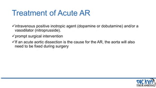
38. Treatment of Acute AR
intravenous positive inotropic agent (dopamine or dobutamine) and/or a
vasodilator (nitroprusside).
prompt surgical intervention
If an acute aortic dissection is the cause for the AR, the aorta will also
need to be fixed during surgery
40. Overview
Backflow of blood from the LV to the LA during systole
Mild (physiological) MR can be seen in normal individuals.
For clinical purposes, MR is classified as
primary and secondary (or functional) MR
o Ischemic MR is subset of secondary MR
o Primary and secondary MR are two distinctly d/t disease conditions, with
d/t pathophysiology, outcomes, and management considerations.
Acute vs Chronic MR
Based on leaflet motion
43. Pathophysiology of chronic
primary MR
Ventricular compensatory
Volume Overload
o Frank-Starling: Increased preload --> LV Dilatation --> increased
stroke volume
o Initially -- Increased LV Ejection Fraction --> increased stroke volume
o (Eventually -- LV systolic function deteriorates and CHF ensues)
Myocardial contractility
Left Atrial Compliance
o Acute: rapidly increasing LA/PV pressure….>pulmonary edema
o Chronic: slow enlargement of the LA with low pressures
47. The severity of MR and the ratio of forward cardiac flow and
backward flow are determined by:
1) the size of the mitral orifice during regurgitation
2) the SVR opposing forward flow from the ventricle
3) the compliance of the left atrium
4) the systolic pressure gradient between the LV and the LA
5) the duration of regurgitation during systole (not all regurgitation is holo-
systolic)
48. Effect of MR on LA pressure depends on LA Compliance
ACUTE MR (non-compliant LA) CHRONIC MR (compliant LA)
Normal LA size Dilated LA
Increased LA pressure LA pressure normal or slow increase
"V" waves on PCW tracing Absence of V waves
Pulmonary Edema Low output state
49. Clinical features
Until late asymptomatic
Acute MR: Dyspnea (pulmonary venous congestion)
Chronic MR: weakness, fatigue and low output state
Palpitation (atrial fibrillation, increased stroke volume)
RV failure (less common than with MS)
Systemic embolization { stroke , ischemic limb etc.. }
51. Diagnosis and Evaluation
• ECHO:
• Cause, underlying disease, Chamber size, severity of regurgitation
• Jet morphology… mechanism of MR (opposite from the most significant anatomic
lesion) except in functional MR
• Quantitative measurements
• TEE
• when TTE images are inadequate
• Whether MVR or clipping is feasible or MVR is necessary
• Detailed anatomy of MV
• 3D Echo
• directly demonstrates pathology (surface rendering)
• Multiple images--- localization of the pathology
• Stress echo
• Can be used to establish the presence of symptoms in patients with chronic primary
MR and exercise tolerance
58. Other Diagnostic Evaluation
Modalities :
ECG: LAE & Afib , LVH (1/3) with severe
MR, RVH (15%)
CXR: Cardiomegaly LV enlargement +
LA enlargement, Calcification (dense C
shaped opacity)
Combined MS + MR…
Cardiac Magnetic Resonance
Imaging.
Accurate measurement of regurgitant
flow
The most accurate non invasive
testing (LVSEDV,ESV and mass)
⁻ indicated in chronic primary MR to
assess LV and RV volumes,
function or MR severity
Cardiac Computed
Tomography:
Sizing mitral annulus and quantifying
degree of annular calcification
particularly useful for planning MVR
Left Ventricular Angiography.
• when discrepancy b/n
noninvasive testing and clinical
symptoms
• Assess Severity of MR, LV
function, need for surgery
• Qualitative but clinically useful
estimate of severity of MR
59. Disease course
Natural history of Chronic primary MR is highly variable and depends on:
oThe volume of regurgitation
o State of the myocardium
o Cause of underlying disorder.
o Asymptomatic mild MR - stable for many years
o Asymptomatic severe MR- progression to symptoms, LV dysfunction,
pulmonary HTN, or AF is 30-40% at 5 years.
60. cont….
5 year survival is 30% in patients who are candidates for operation,
presumably b/c of symptoms, but who declined surgery
In Severe MR due to flail leaflets
₋ annual mortality without surgery is 3%
₋ at 20 years 60%
₋ it is high in those with LV Dysfunction (LVEF of 60% or less)
66. Management of MR
Medical management
In patients with primary MR, there is no convincing evidence
that vasodilator therapy reduces MR severity.
However, GDMT for LV systolic dysfunction or systemic HTN
should be implemented as in any patient with these
conditions.
69. Mitral valve Surgery
Only effective treatment is valve repair/replacement
Optimal timing is determined by:
• Presence/absence of symptoms
• Functional state of ventricle
• Feasibility of valve repair
• Presence of Afib/PHTN
• Presence/expectation of patient
70. cont….
Surgical mortality depends on the patient's clinical and hemodynamic
status particularly:
₋ LV function
₋ age
₋ presence of comorbid conditions such as renal, hepatic, or pulmonary
disease
₋ the skill and experience of the surgical team
71. MV Repair vs. Replacement
Valve replacement:
oMortality 2-7%
oAnti-coagulation
oDecreased LVEF
Prosthesis related complications
Mechanical prosthetic valve
dysfunction/ thrombosis
Valve repair
oMortality 2-3%
oNo anticoagulation (unless Afib)
oPreservation of LVEF
Valve repair always preferable
Feasible in 70-90% of patients
High volume surgeon (20
procedures/yr)
72. Indication for surgery
Asymptomatic patients :
LV dysfunction (EF <60% and /or LV end-diastolic diameter of 40 mm)
AF or pulmonary hypertension
Symptomatic patients :
Patients with severe primary MR and moderate or severe symptoms
(NYHA Class II, III, and IV) should generally be considered for surgery.
74. MV Repair vs. Replacement
Repair of primary degenerative MR most often is successful in:
1) children and adolescent with pliable valves
2) adults with MR secondary to MVP
3) cases with annular dilatation
4) cases with chordal rupture
5) cases with perforation of a mitral leaflet caused by infective
endocarditis
75. Percutaneous mitral valve repair
Mitra Clip is safe (Everest I) but less effective than surgical repair (Everest II)
Residual MR by creating 2 regurgitant orifice – similar to Alfieri procedure
Reduce symptoms by reducing MR, reverse LV remodeling
For patients:
o with chronic severe MR with symptoms NYHA 3-4 despite medical therapy for
HF and are not candidate for surgery
o Favorable anatomy for repair procedure
o Reasonable life expectancy
76. Surgical results
Operative mortality is 1-9% in many centers
Mortality was significantly lower in the repair than replacement
patients (1.4% vs 5.4% in 2007-10)
The mortality rate in mitral valve surgery with CABG is
6.2%... 25% in pts with LV dysfunction
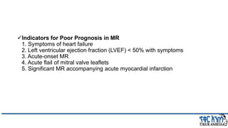
77. Indicators for Poor Prognosis in MR
1. Symptoms of heart failure
2. Left ventricular ejection fraction (LVEF) < 50% with symptoms
3. Acute-onset MR
4. Acute flail of mitral valve leaflets
5. Significant MR accompanying acute myocardial infarction
78. Surgical result
A large proportion of operative
survivors after MVR or repair exhibit:
• improved clinical status
• quality of life
• Exercise tolerance
If operation done before development
of serious symptoms or marked LV
dysfunction:
• Severe PHTN is reduced
• LVEDV and LV mass decrease
• coronary flow reserve increases.
• Depressed contractile function
improves
79. Treatment of Chronic Secondary
MR
Medical Management
GDMT for HF with reduced LVEF in patients with severe secondary MR
(I-A)
Treatment should be provided, in conjunction with a cardiology expert,
in the management of HF (I-C)
80. Surgical Treatment in chronic secondary MR
If patient has persistent symptoms despite optimal medical therapy
Reasonable if severe secondary MR undergoing surgery for CABG
but Several randomized trials have provided conflicting data on concomitant
MV repair in pts undergoing CABG.
POINT Trial
RIME Trial
CTSN Trial
Transcatheter Treatment of secondary MR
COAPT trial Vs MITRA-FR
83. Eur Heart J Cardiovasc Imaging, Volume 20, Issue 6, June 2019, Pages 620–624, https://doi.org/10.1093/ehjci/jez073
The content of this slide may be subject to copyright: please see the slide notes for details.
Utility vs. futility of MitraClip procedure according to severity of MR
and LV systolic dysfunction. EROA, ...
84. MV repair vs replacement in secondary
chronic MR
87. Treatment of Acute MR
Medical Treatment
Afterload reduction -IV nitroprusside and add inotropic agent (I. e
dobutamine) if hypotension
intra-aortic balloon pump
Emergent Surgery - mitral valve repair or replacement
Surgical mortality rates are higher in patients with:
acute MR and refractory HF (NYHA Class IV)
those with prosthetic valve dysfunction
those with active infective endocarditis
90. Pathophysiology TR
The RA and RV end-diastolic pressures often are elevated in TR whether
the condition is caused by:
oorganic disease of the tricuspid valve
o is secondary to RV systolic overload.
Determination of the pulmonary artery (or RV) systolic pressure may be
helpful in deciding whether the TR is primary or secondary to RV dilation
94. Clinical presentation of TR
In absence of PHTN or RV failure, TR generally is well tolerated.
Manifested with of right-sided HF: reduced CO, ascites, painful congestive
hepatomegaly, and massive edema
95. Physical Examination
The RV impulse is hyperdynamic and thrusting in quality.
High pitch blowing holosystolic murmur varying with respiration(carvallo
sign) in xiphoid area
Large pulsations in neck, pulsatile enlarged liver, widespread edema, RV
S3(increase with respiration)
In TR 2ndary to PHTN
P2 is accentuated
systolic murmur usually is high pitched, pansystolic
loudest in the 4th intercostal space in the parasternal region
96. Investigation
ECHO:
to estimate the severity of TR and assess pulmonary artery pressure (PAP) and RV
function, RV and RA chamber size , mass, valve anatomy and motion
ECG:
nonspecific or Incomplete right bundle branch block, Q waves in lead V1, and AF are
often found.
CXR:
marked cardiomegaly, and the right atrium is prominent, distention of the azygos vein
and the presence of a pleural
effusion.
Cardiac Magnetic Resonance Imaging:
for determining the 3 D geometric relationships between the right ventricle and the
tricuspid annulus and leaflets in patients with functional TR
98. Management
Medical
Diuretic therapy treats the systemic congestion in patients with severe symptomatic TR (2a)
In patients with secondary TR, treatment of the underlying primary cause may decrease the
severity of the TR (2a)
Surgery:
• Severe functional TR may be treated by annuloplasty or valve replacement.
• Severe TR due to intrinsic tricuspid valve disease requires valve replacement.
• Tricuspid valve repair accounts for 73% of tricuspid valve operations.
• For TVR, bio prostheses are increasingly being used and now account for 46% of
TVRs.
99. Treatment…
• Treatment of secondary TR is targeted at pulmonary
hypertension or myocardial disease.
• Outcomes of patients with severe primary TR are poor with
medical management.
• Surgical treatment is performed for selected patients with
• TR at the time of surgery for left-sided valve lesions to treat severe TR (Stages C and
D) and
• to prevent later development of severe TR in patients with progressive TR (Stage B).
• Severe isolated TR
• Mild TR with annular dilatation
100. Treatment…
• There is renewed interest in earlier surgery for patients with
severe isolated TR before the onset of severe RV dysfunction or
end-organ damage.
• This interest is attributable to
• 1) an increasing no. of patients presenting with RHF from isolated TR
• 2) more advanced surgical techniques, and
• 3) better selection processes, resulting in a lower operative risk with
documented improvement in symptoms
• There is growing interest in the development of catheter-based
therapies for these patients with severe isolated TR.
103. Pulmonary regurgitation
Most common cause is ring dilatation due to pulmonary hypertension, or
dilatation of the pulmonary artery secondary to a connective tissue disorder.
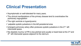
104. Clinical Presentation
Asymptomatic or well-tolerated for many years.
The clinical manifestations of the primary disease tend to overshadow the
pulmonary regurgitation
The right ventricle is hyperdynamic
palpable systolic pulsations in the left parasternal area
enlarged pulmonary artery often produces systolic pulsations in the 2nd left
intercostal space.
the diastolic murmur of PR is low-pitched and usually is heard best at the 3rd and
4th left intercostal spaces adjacent to the sternum.
105. Cont….
Electrocardiography.
o an rSr (or rsR) configuration in the right precordial leads.
o RV hypertrophy.
Radiography.
o Both the pulmonary artery and right ventricle are usually enlarged, but these
signs are nonspecific.
Cardiac Magnetic Resonance Imaging.
o assess pulmonic valve anatomy
o recognize any obstruction above or below the valve
o measure pulmonary artery dilation
o quantify PR severity.
o evaluation of RV dilation and systolic function
106. Echocardiography
Right chamber Size , RV hypertrophy, RV function , abnormal motion of
septum and motion of pulmonic valve.
Doppler echo - is extremely accurate in detecting PR and in helping
estimate its severity