18 basics of pediatric airway anatomy, physiology and management
- 1. Basics of Pediatric Airway Anatomy, Physiology and Management Christine Mai, MD Claudine Mansour, MD Faculty Advisor: Ruth Padilla, MD Boston University Medical Center Department of Anesthesiology
- 2. The Pediatric Airway Introduction Normal Anatomy Physiology Airway evaluation Management of normal vs. abnormal airway Difficult airway
- 3. Introduction Almost all of pediatric codes are due to respiratory origin 80% of pediatric cardiopulmonary arrest are primarily due to respiratory distress Majority of cardiopulmonary arrest occur at <1 year old 1990 Closed Claim Project by ASA Respiratory events are the largest class of injury (34%) More common in children than adults 92% of claims occurred between 1975-1985 before continuous pulsoximetry and capnography (Brain damage and death in 85% of cases) With continuous O 2 sat and ETCO 2 monitoring after 1990s, decrease in brain damage and death (56% 1970s to 31% 1990s)
- 4. Normal Pediatric Airway Anatomy Larynx composed of hyoid bone and a series of cartilages Single: thyroid, cricoid, epiglottis Paired: arytenoids, corniculates, and cuneiform
- 5. Pediatric Anatomy cont. Laryngeal folds consist of: Paired aryepiglottic folds extend from epiglottis posteriorly to superior surface of arytenoids Paired vestibular folds (false vocal cords) extend from thyroid cartilage posteriorly to superior surface of arytenoids Paired vocal folds (true vocal cords) extend from posterior surface of thyroid plate to anterior part of arytenoids Interarytenoid fold bridging the arytenoid cartilages Thyrohyoid fold extend from hyoid bone to thyroid cartilage Sensory Innervation: Recurrent Laryngeal Nerve-supraglottic larynx Internal Branch of Superior Laryngeal Nerve-infraglottic larynx Motor Innervation: External branch of Superior Laryngeal Nerve-cricothyroid muscle Recurrent Laryngeal Nerve-all other laryngeal muscles Blood Supply Laryngeal branches of the superior and inferior thyroid arteries
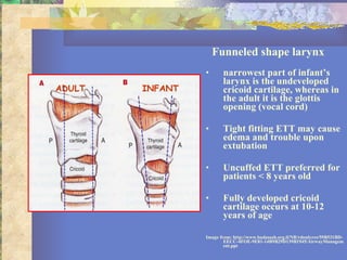
- 6. 5 Differences between Pediatric and Adult Airway More rostral larynx Relatively larger tongue Angled vocal cords Differently shaped epiglottis Funneled shaped larynx-narrowest part of pediatric airway is cricoid cartilage
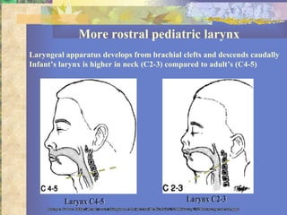
- 7. More rostral pediatric larynx Laryngeal apparatus develops from brachial clefts and descends caudally Infant’s larynx is higher in neck (C2-3) compared to adult’s (C4-5) Larynx C4-5 Larynx C2-3 Image from: http://depts.washington.edu/pccm/Pediatric%20Airway%20management.ppt
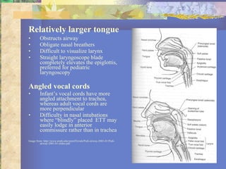
- 8. Relatively larger tongue Obstructs airway Obligate nasal breathers Difficult to visualize larynx Straight laryngoscope blade completely elevates the epiglottis, preferred for pediatric laryngoscopy Angled vocal cords Infant’s vocal cords have more angled attachment to trachea, whereas adult vocal cords are more perpendicular Difficulty in nasal intubations where “blindly” placed ETT may easily lodge in anterior commissure rather than in trachea Image from: http://www.utmb.edu/otoref/Grnds/Pedi-airway-2001-01/Pedi-airway-2001-01-slides.pdf
- 9. Differently shaped epiglottis Adult epiglottis broader, axis parallel to trachea Infant epiglottis ohmega ( Ώ ) shaped and angled away from axis of trachea More difficult to lift an infant’s epiglottis with laryngoscope blade
- 10. Funneled shape larynx narrowest part of infant’s larynx is the undeveloped cricoid cartilage, whereas in the adult it is the glottis opening (vocal cord) Tight fitting ETT may cause edema and trouble upon extubation Uncuffed ETT preferred for patients < 8 years old Fully developed cricoid cartilage occurs at 10-12 years of age Image from: http://www.hadassah.org.il/NR/rdonlyres/59B531BD-EECC-4FOE-9E81-14B9B29D139B1945/AirwayManagement.ppt INFANT ADULT
- 11. Pediatric Respiratory Physiology Extrauterine life not possible until 24-25 weeks of gestation Two types of pulmonary epithelial cells: Type I and Type II pneumocytes Type I pneumocytes are flat and form tight junctions that interconnect the interstitium Type II pneumocytes are more numerous, resistant to oxygen toxicity, and are capable of cell division to produce Type I pneumocytes Pulmonary surfactant produced by Type II pneumocytes at 24 wks GA Sufficient pulmonary surfactant present after 35 wks GA Premature infants prone to respiratory distress syndrome (RDS) because of insufficient surfactant Betamethasone can be given to pregnant mothers at 24-35wks GA to accelerate fetal surfactant production
- 12. Pediatric Respiratory Physiology cont. Work of breathing for each kilogram of body weight is similar in infants and adult Oxygen consumption of infant (6 ml/kg/min) is twice that of an adult (3 ml/kg/min) Greater oxygen consumption = increased respiratory rate Tidal volume is relatively fixed due to anatomic structure Minute alveolar ventilation is more dependent on increased respiratory rate than on tidal volume Lack Type I muscle fibers, fatigue more easily FRC of an awake infant is similar to an adult when normalized to body weight Ratio of alveolar minute ventilation to FRC is doubled, under circumstances of hypoxia, apnea or under anesthesia, the infant’s FRC is diminished and desaturation occurs more precipitously
- 13. Physiology: Effect Of Edema Poiseuille’s law R = 8nl/ π r 4 If radius is halved, resistance increases 16 x Image from: http://www.hadassah.org.il/NR/rdonlyres/59B531BD-EECC-4FOE-9E81-14B9B29D139B1945/AirwayManagement.ppt
- 14. Normal Inspiration and Expiration turbulence Inspiration Expiration Image from: http://www.hadassah.org.il/NR/rdonlyres/59B531BD-EECC-4FOE-9E81-14B9B29D139B1945/AirwayManagement.ppt
- 15. Obstructed Airways turbulence & wheezing Extrathoracic Upper Airway Obstruction Intrathoracic Lower Airway Obstruction Epiglottitis, laryngotracheobronchitis, foreign body Asthma, bronchiolitis Image from: http://www.hadassah.org.il/NR/rdonlyres/59B531BD-EECC-4FOE-9E81-14B9B29D139B1945/AirwayManagement.ppt
- 16. URI predisposes to coughing, laryngospasm, bronchospasm, desat during anesthesia Snoring or noisy breathing (adenoidal hypertrophy, upper airway obstruction, OSA) Chronic cough (subglottic stenosis, previous tracheoesohageal fistula repair) Productive cough (bronchitis, pneumonia) Sudden onset of new cough (foreign body aspiration) Inspiratory stridor (macroglossia, laryngeal web, laryngomalacia, extrathoracic foreign body) Hoarse voice (laryngitis, vocal cord palsy, papillomatosis) Asthma and bronchodilator therapy (bronchospasm) Repeated pneumonias (GERD, CF, bronchiectasis, tracheoesophageal fistula, immune suppression, congenital heart disease) History of foreign body aspiration Previous anesthetic problems (difficulty intubation/extubation or difficulty with mask ventilation) Atopy, allergy (increased airway reactivity) History of congenital syndrome (Pierre Robin Sequence, Treacher Collins, Klippel-Feil, Down’s Syndrome, Choanal atresia) Environmental: smokers Airway Evaluation Medical History
- 17. Signs of Impending Respiratory Failure Increase work of breathing Tachypnea/tachycardia Nasal flaring Drooling Grunting Wheezing Stridor Head bobbing Use of accessory muscles/retraction of muscles Cyanosis despite O 2 Irregular breathing/apnea Altered consciousness/agitation Inability to lie down Diaphoresis
- 18. Airway Evaluation Physical Exam Facial expression Nasal flaring Mouth breathing Drooling Color of mucous membranes Retraction of suprasternal, intercostal or subcostal Respiratory rate Voice change Mouth opening Size of mouth Mallampati Loose/missing teeth Size and configuration of palate Size and configuration of mandible Location of larynx Presence of stridor (inspiratory/expiratory) Baseline O2 saturation Global appearance (congenital anomalies) Body habitus
- 19. Diagnostic Testing Laboratory and radiographic evaluation extremely helpful with pathologic airway AP and lateral films and fluoroscopy may show site and cause of upper airway obstruction MRI/CT more reliable for evaluating neck masses, congenital anomalies of the lower airway and vascular system Perform radiograph exam only when there is no immediate threat to the child’s safety and in the presence of skilled personnel with appropriate equipment to manage the airway Intubation must not be postponed to obtain radiographic diagnosis when the patient is severely compromised. Blood gases are helpful in assessing the degree of physiologic compromise; however, performing an arterial puncture on a stressed child may aggravate the underlying airway obstruction
- 20. Airway Management: Normal Airway Challenging because of unique anatomy and physiology Goals: protect the airway, adequately ventilate, and adequately oxygenate Failure to perform any ONE of these tasks will result in respiratory failure Positioning is key!
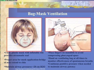
- 21. Bag-Mask Ventilation Clear, plastic mask with inflatable rim provides atraumatic seal Proper area for mask application-bridge of nose extend to chin Maintain airway pressures <20 cm H2O Place fingers on mandible to avoid compressing pharyngeal space Hand on ventilating bag at all times to monitor effectiveness of spontaneous breaths Continous postitive pressure when needed to maintain airway patency Image from: http://www.hadassah.org.il/NR/rdonlyres/59B531BD-EECC-4FOE-9E81-14B9B29D139B1945/AirwayManagement.ppt
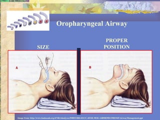
- 22. Oropharyngeal Airway SIZE PROPER POSITION Image from: http://www.hadassah.org.il/NR/rdonlyres/59B531BD-EECC-4FOE-9E81-14B9B29D139B1945/AirwayManagement.ppt
- 23. Oropharyngeal Airway Placement Wrong size: Too long Correct Size Wrong size: Too short Image from: http://depts.Washington.edu/pccm/Pediatric%20Airway%20management.ppt
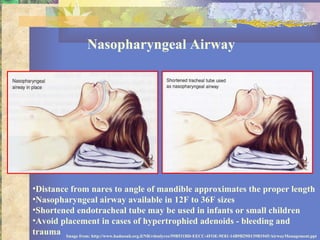
- 24. Nasopharyngeal Airway Distance from nares to angle of mandible approximates the proper length Nasopharyngeal airway available in 12F to 36F sizes Shortened endotracheal tube may be used in infants or small children Avoid placement in cases of hypertrophied adenoids - bleeding and trauma Image from: http://www.hadassah.org.il/NR/rdonlyres/59B531BD-EECC-4FOE-9E81-14B9B29D139B1945/AirwayManagement.ppt
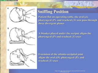
- 25. Sniffing Position Image from: http://depts.Washington.edu/pccm/Pediatric%20Airway%20management.ppt Patient flat on operating table, the oral (o), pharyngeal (P), and tracheal (T) axis pass through three divergent planes A blanket placed under the occiput aligns the pharyngeal (P) and tracheal (T) axes Extension of the atlanto-occipital joint aligns the oral (O), pharyngeal (P), and tracheal (T) axes
- 26. Selection of laryngoscope blade: Miller vs. Macintosh Miller blade is preferred for infants and younger children Facilitates lifting of the epiglottis and exposing the glottic opening Care must be taken to avoid using the blade as a fulcrum with pressure on the teeth and gums Macintosh blades are generally used in older children Blade size dependent on body mass of the patient and the preference of the anesthesiologist
- 27. Endotracheal Tube New AHA Formulas: Uncuffed ETT: (age in years/4) + 4 Cuffed ETT: (age in years/4) +3 ETT depth (lip): ETT size x 3 Age Wt ETT(mm ID) Length(cm ) Preterm 1 kg 2.5 6 1-2.5 kg 3.0 7-9 Neonate-6mo 3.0-3.5 10 6 mo-1 3.5-4.0 11 1-2 yrs 4.0-5.0 12
- 28. Complications of Endotracheal Intubation Postintubation Croup Incidence 0.1-1% Risk factors: large ETT, change in patient position introp, patient position other than supine, multiple attempts at intubation, traumatic intubation, pts ages 1-4, surgery >1hr, coughing on ETT, URI, h/o croup Tx: humidified mist, nebulized racemic epinephrine, steroid Laryngotracheal (Subglottic) Stenosis Occurs in 90% of prolonged endotracheal intubation Lower incidence in preterm infants and neonates due to relative immaturity of cricoid cartilage Pathogenesis: ischemic injury secondary to lateral wall pressure from ETT edema, necrosis, and ulceration of mucosa, infx Granulation tissues form within 48hrs leads to scarring and stenosis
- 29. Cuff vs Uncuffed Endotracheal Tube Controversial issue Traditionally, uncuffed ETT recommended in children < 8 yrs old to avoid post-extubation stridor and subglottic stenosis Arguments against cuffed ETT: smaller size increases airway resistance, increase work of breathing, poorly designed for pediatric pts, need to keep cuff pressure < 25 cm H2O Arguments against uncuffed ETT: more tube changes for long-term intubation, leak of anesthetic agent into environment, require more fresh gas flow > 2L/min, higher risk for aspiration -Concluding Recommendations- For “short” cases when ETT size >4.0, choice of cuff vs uncuffed probably does not matter Cuffed ETT preferable in cases of: high risk of aspiration (ie. Bowel obstruction), low lung compliance (ie. ARDS, pneumoperitoneum, CO2 insufflation of the thorax, CABG), precise control of ventilation and pCO2 (ie. increased intracranial pressure, single ventricle physiology) Golden, S. “Cuffed vs. Uncuffed Endotracheal tubes in children: A review” Society for Pediatric Anesthesia. Winter 2005 edition.
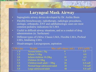
- 30. Laryngeal Mask Airway Supraglottic airway device developed by Dr. Archie Brain Flexible bronchoscopy, radiotherapy, radiologic procedures, urologic, orthopedic, ENT and ophthalmologic cases are most common pediatric indications for LMA Useful in difficult airway situations, and as a conduit of drug administration (ie. Surfactant) Different types of LMAs: Classic LMA, Flexible LMA, ProSeal LMA, Intubating LMA Disadvantages: Laryngospasm, aspiration LMA size Weight Max cuff volume (mL) ETT (mID) 1 .0 Neonate/Infants ≤ 5kg 4 3.5 1.5 Infants 5-10kg 7 4.0 2.0 Infants/children 10-20kg 10 4.5 2.5 Children 20-30kg 14 5.0 3.0 Children/small adult > 30kg 20 6.0 cuff 4.0 Normal/large adolescent/adult 30 7.0 cuff 5.0 Large adolescent/adult 40 8.0 cuff
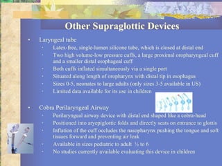
- 31. Other Supraglottic Devices Laryngeal tube Latex-free, single-lumen silicone tube, which is closed at distal end Two high volume-low pressure cuffs, a large proximal oropharyngeal cuff and a smaller distal esophageal cuff Both cuffs inflated simultaneously via a single port Situated along length of oropharynx with distal tip in esophagus Sizes 0-5, neonates to large adults (only sizes 3-5 available in US) Limited data available for its use in children Cobra Perilaryngeal Airway Perilaryngeal airway device with distal end shaped like a cobra-head Positioned into aryepiglottic folds and directly seats on entrance to glottis Inflation of the cuff occludes the nasopharynx pushing the tongue and soft tissues forward and preventing air leak Available in sizes pediatric to adult ½ to 6 No studies currently available evaluating this device in children
- 32. Difficult Airway Management Techniques Rigid bronchoscopy Flexible bronchoscopy Direct laryngoscopy Intubating LMA Lighted stylet Bullardscope Fiberoptic intubation Surgical airway
- 33. Airway Management Classification of Abnormal Pediatric Airway Congenital Neck Masses (Dermoid cysts, cystic teratomas, cystic hygroma, lymphangiomas, neurofibroma, lymphoma, hemangioma) Congenital Anomalies (Choanal atresia,tracheoesophageal fistula, tracheomalacia, laryngomalacia, laryngeal stenosis, laryngeal web, vascular ring, tracheal stenosis) Congenital Syndromes (Pierre Robin Syndrome, Treacher Collin, Turner, Down’s, Goldenhar , Apert, Achondroplasia, Hallermann-Streiff, Crouzan) Inflammatory (Epiglottitis, acute tonsillitis, peritonsillar abscess,retropharyngeal abscess, laryngotracheobronchitis,bacterial tracheitis,adenoidal hypertrophy,nasal congestion, juvenile rheumatoid arthritis) Traumatic/Foreign Body (burn,laceration,lymphatic/venous obstruction,fractures/dislocation, inhalational injury, postintubation croup (edema),swelling of uvula Metabolic (Congenital hypothyroidism, mucopolysaccharidosis, Beckwith-Wiedemann Syndrome,glycogen storage disease, hypocalcemia laryngospasm)
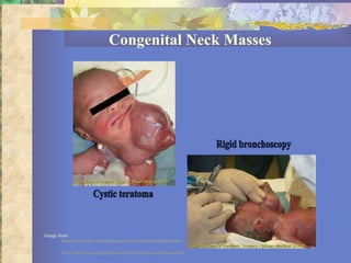
- 34. Congenital Neck Masses Image from: http://bms.brown.edu/pedisurg/fetal/seminar/imagebank.html http://bms.brown.edu/pedisurg/Brown/IBImages/Teratoma/BronchospyTeratoma.html Cystic teratoma Rigid bronchoscopy
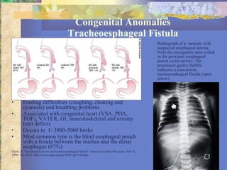
- 35. Congenital Anomalies Tracheoesphageal Fistula Feeding difficulties (coughing, choking and cyanosis) and breathing problems Associated with congenital heart (VSA, PDA, TOF), VATER, GI, musculoskeletal and urinary tract defects Occurs in 1/ 3000-5000 births Most common type is the blind esophageal pouch with a fistula between the trachea and the distal esophagus (87%) Clark, D. “Esophageal atresia and tracheoesophageal fistula” American Family Physician. Feb 15, 1999. Vol 59(4) http://www.aafp.org/afp/99021ap/910.htlm Radiograph of a neonate with suspected esophageal atresia. Note the nasogastric tube coiled in the proximal esophageal pouch (solid arrow). The prominent gastric bubble indicates a concurrent tracheoesphageal fistula (open arrow)
- 36. Congenital Anomalies Choanal Atresia Complete nasal obstruction of the newborn Occurs in 0.82/10 000 births During inspiration, tongue pulled to palate, obstructs oral airway Unilateral nare (right>left) Bilateral choanal atresia is airway emergency Death by asphyxia Associated with other congenital defects Tewfik, T. “Choanal atresia” emedicine.com http://www.emedicine.com/ent/topic330.htm
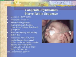
- 37. Congenital Syndromes Pierre Robin Sequence Occurs in 1/8500 births Autosomal recessive Mandibular hypoplasia, micrognathia, cleft palate, retraction of inferior dental arch, glossptosis Severe respiratory and feeding difficulties Associated with OSA, otitis media, hearing loss, speech defect, ocular anomalies, cardiac defects, musculoskeletal (syndactyly, club feet), CNS delay, GU defects) Tewfik, T. “Pierre Robin Syndrome” emedicine.com http://www.emedicine.com/ent/topic150.htm
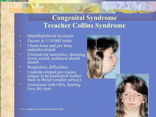
- 38. Congenital Syndrome Treacher Collins Syndrome Mandibulofacial dysotosis Occurs in 1/10 000 births Cheek bone and jaw bone underdeveloped External ear anamolies, drooping lower eyelid, unilateral absent thumb Respiratory difficulties Underdeveloped jaw causes tongue to be positioned further back in throat (smaller airway) Associated with OSA, hearing loss, dry eyes www.ccakids.com/syndrome/treacher.pdf
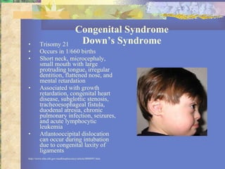
- 39. Congenital Syndrome Down’s Syndrome Trisomy 21 Occurs in 1/660 births Short neck, microcephaly, small mouth with large protruding tongue, irregular dentition, flattened nose, and mental retardation Associated with growth retardation, congenital heart disease, subglottic stenosis, tracheoesophageal fistula, duodenal atresia, chronic pulmonary infection, seizures, and acute lymphocytic leukemia Atlantooccipital dislocation can occur during intubation due to congenital laxity of ligaments http://www.nlm.nih.gov/medlineplus/ency/article/0000997.htm
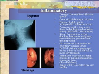
- 40. Inflammatory Etiology: Haemophilus influenzae type B Occurs in children ages 2-6 years Disease of adults due to widespread H. influenza vaccine Progresses rapidly from a sore throat to dysphagia and complete airway obstruction (within hours) Signs of obstruction: stridor, drooling, hoarseness, tachypnea, chest retraction, preference for upright position OR intubation/ENT present for emergency surgical airway Do NOT perform laryngoscopy before induction of anesthesia to avoid laryngospasm Inhalational induction in sitting position to maintain spontaneous respiratory drive (Sevo/Halothane) Range of ETT one-half to one size smaller Epiglottitis Thumb sign
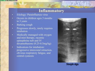
- 41. Inflammatory Etiology: Parainfluenza virus Occurs in children ages 3 months to 3 years Barking cough Progresses slowly, rarely requires intubation Medically managed with oxygen and mist therapy, racemic epinephrine neb and IV dexamethasone (0.25-0.5mg/kg) Indications for intubation: progressive intercostal retraction, obvious respiratory fatigue, and central cyanosis Steeple sign
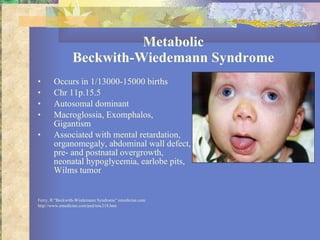
- 42. Metabolic Beckwith-Wiedemann Syndrome Occurs in 1/13000-15000 births Chr 11p.15.5 Autosomal dominant Macroglossia, Exomphalos, Gigantism Associated with mental retardation, organomegaly, abdominal wall defect, pre- and postnatal overgrowth, neonatal hypoglycemia, earlobe pits, Wilms tumor Ferry, R “Beckwith-Wiedemann Syndrome” emedicine.com http://www.emedicine.com/ped/toic218.htm
- 43. Pediatric Difficult Airway Algorithm Rigid bronchoscopy The unexpected difficult pediatric intubation Mask ventilation possible? Succeed Non emergency pathway If mask ventilation becomes inadequate Fail Emergency Pathway Call for help Awaken patient Succeed Succeed Failed Failed Awaken patient Establish definitive airway Surgical airway GA by mask GA by LMA Surgical airway Surgical airway Awake intubation technique Regional anesthesia Abort and regroup Persue other intubation options Change head position Different intubation technique Different technique with rigid laryngoscope LMA as intubation guide Fiberoptic Light wand Retrograde OELM Different blades Stylets Retromolar approach Persue emergency oxygenation/ventilation options Two person mask ventilation Percutaneous cricothyrotomy LMA
Editor's Notes
- 9
- 16

