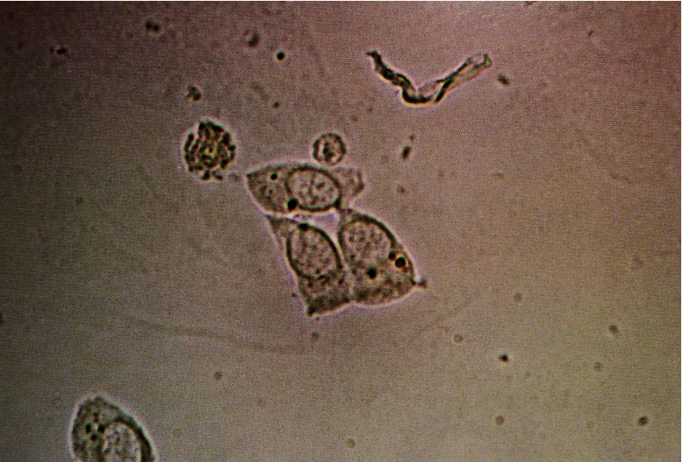
Three renal epithelial cells, whose nuclei are approximately the same size as an adjacent neutrophil. Renal epithelial cells are polyhedral and columnar. Their nuclei are often slightly off center and displaced towards the cell base. A microvillus border is evident in the cells shown here
200X
Source: Urinary Sediment: A Textbook Atlas. Haber, Meryl H.
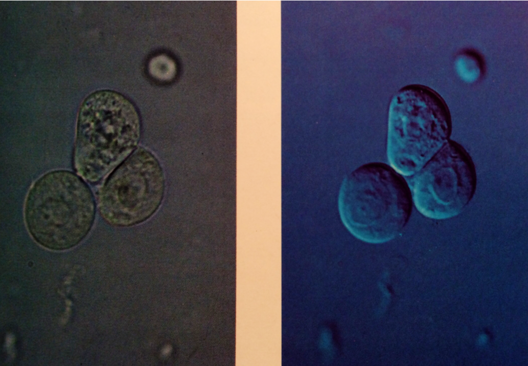
L: Transitional epithelial cells, which are nearly spheric due to absorption of water and have large, central nuclei
320X
R: Transitional epithelial cells in urinary sediment. This relief image clarifies spheric shape of these cells and nuclear detail and is superior to ordinary brightfield microscopy for this purpose
Interference-contrast microscopy, 320X
Source: Urinary Sediment: A Textbook Atlas. Haber, Meryl H.
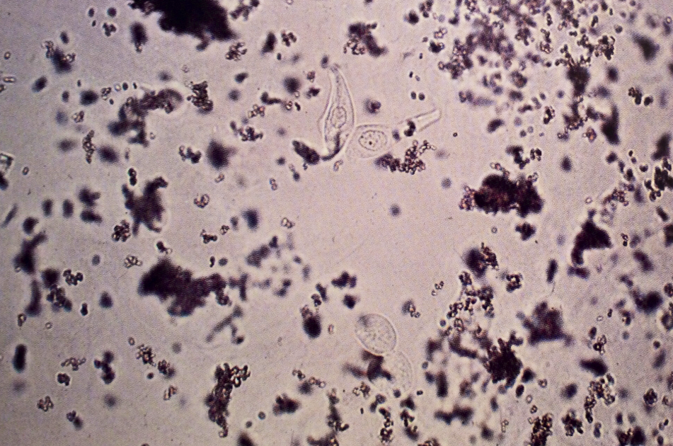
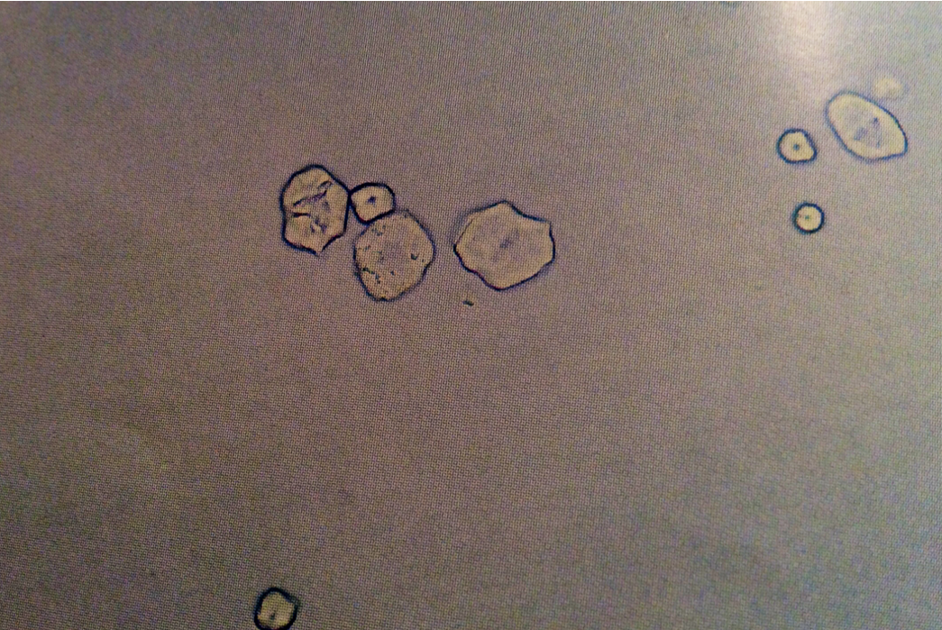
Four transitional epithelial cells (center). Although there is much amorphous crystalline material in the field, the cells are still easily recognized. Two are “tadpole”-shaped, whereas the other two are spheric
160X
Source: Urinary Sediment: A Textbook Atlas. Haber, Meryl H.
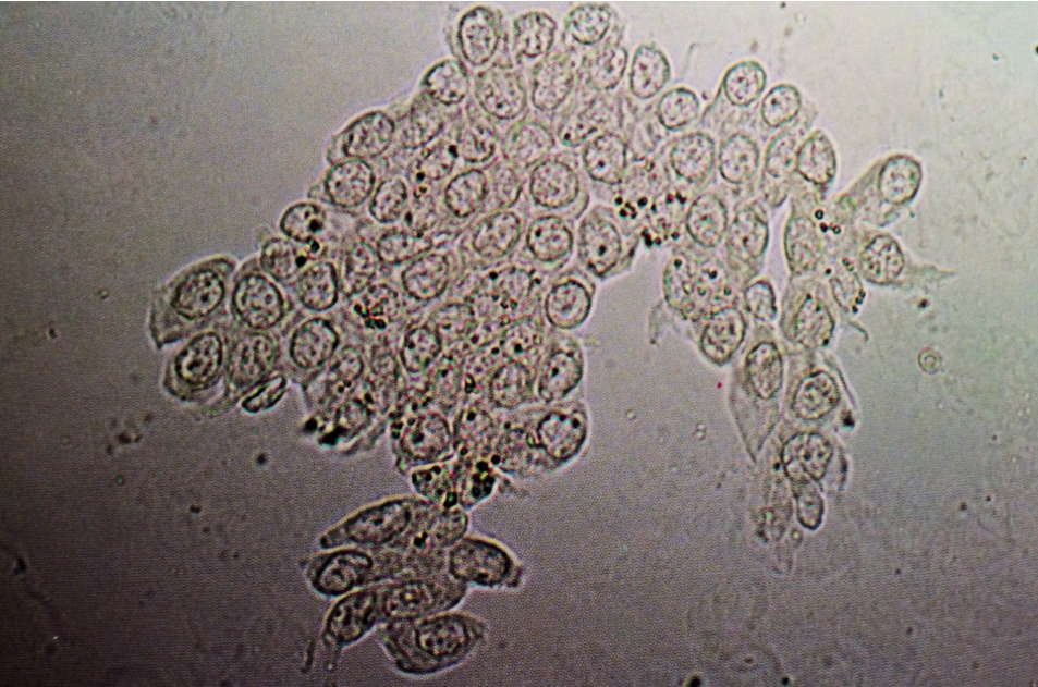
Group of transitional epithelial cells in urine, which are spheric and contain central large nuclei
160X
Source: Urinary Sediment: A Textbook Atlas. Haber, Meryl H.
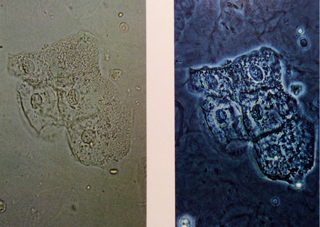
L: Squamous epithelial cells, commonly seen in clumps, but also singly. They are large, flat, and often have finely wrinkled cytoplasm
200X
R: Group of squamous epithelial cells with mucus strands in the background. Here, the difference between phase-contrast and brightfield microscopy is demonstrated
Phase-contrast microscopy, 200X
Source: Urinary Sediment: A Textbook Atlas. Haber, Meryl H.
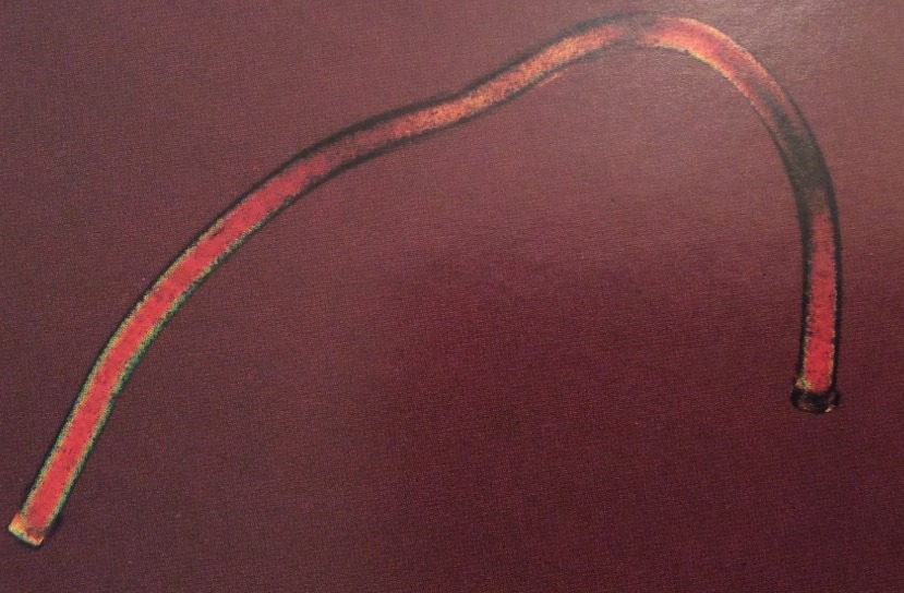
Fiber under polarized light
Only fatty casts will polarize
100X
Source: Urinalysis and Body Fluids: Strasinger, Susan K.
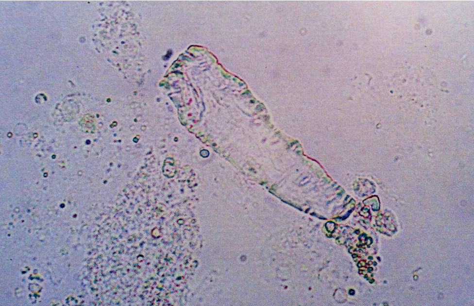
Artifact resembling a waxy cast
Notice lack of typical cast form and refractility
400X
Source: Urinalysis and Body Fluids: Strasinger, Susan K.
Starch granules
Notice the refractility
400X
Source: Urinalysis and Body Fluids: Strasinger, Susan K.
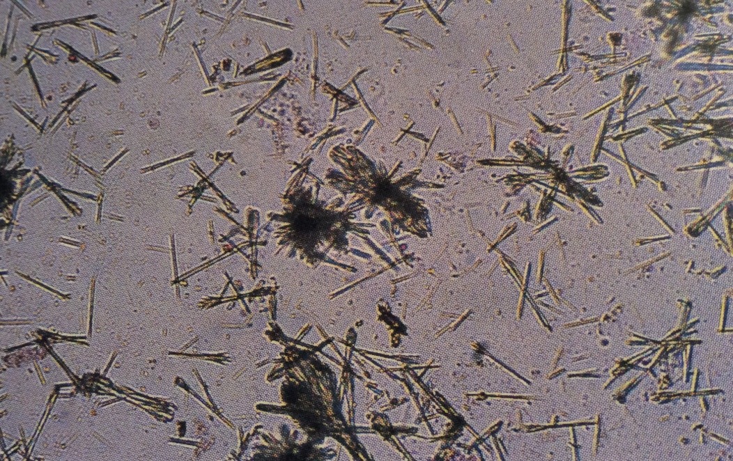
Ampicillin crystals
Bundles of crystals are seen after refrigeration
400X
Source: Urinalysis and Body Fluids: Strasinger, Susan K.
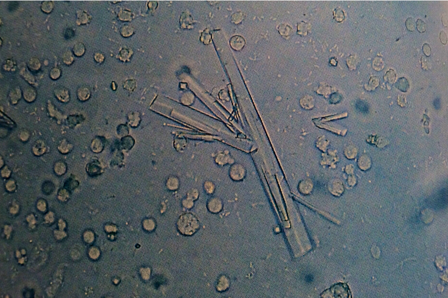
Sulfa crystals, white blood cells and bacteria suggest a urinary tract infection
400X
Source: Urinalysis and Body Fluids: Strasinger, Susan K.
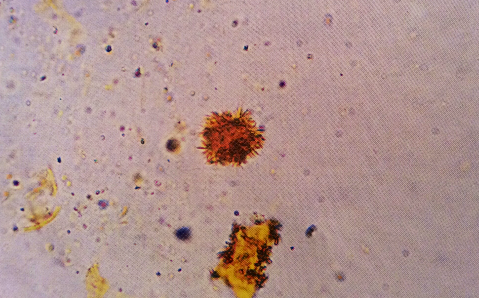
Bilirubin crystals
Notice the classic bright yellow color
400X
Source: Urinalysis and Body Fluids: Strasinger, Susan K.
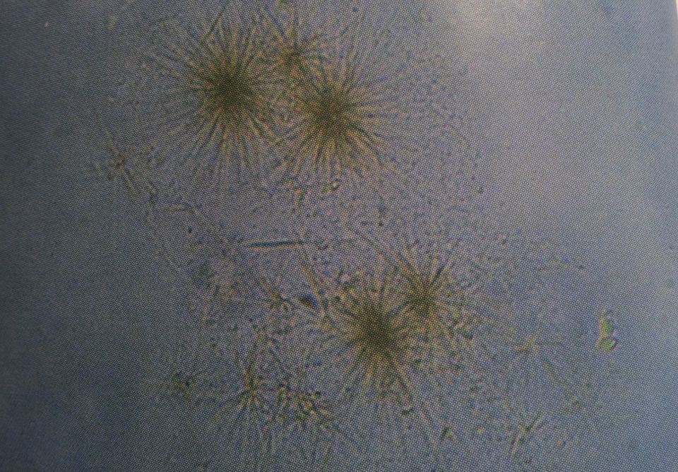
Tyrosine crystals
Yellow color suggests liver disease
400X
Source: Urinalysis and Body Fluids: Strasinger, Susan K.
Cholesterol crystals
Notice notched corners
400X
Source: Urinalysis and Body Fluids: Strasinger, Susan K.
