
Starch granules, relatively regular spherules derived from cornstarch and commonly found in urine
100X
Source: Urinary Sediment: A Textbook Atlas. Haber, Meryl H.
 16th April 2015
 Notes: 3


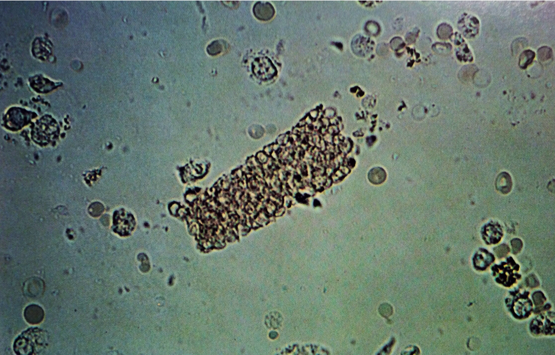
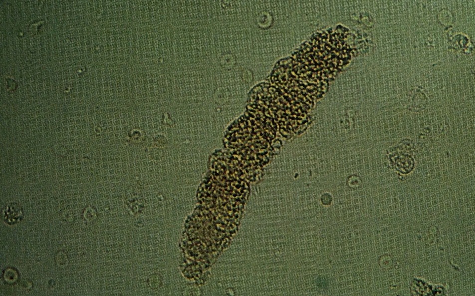
Epithelial cell cast. Several well circumscribed and intact tubular epithelial cells, imbedded in a hyaline cast matrix, are easily identified as to type
160X
Source: Urinary Sediment: A Textbook Atlas. Haber, Meryl H.

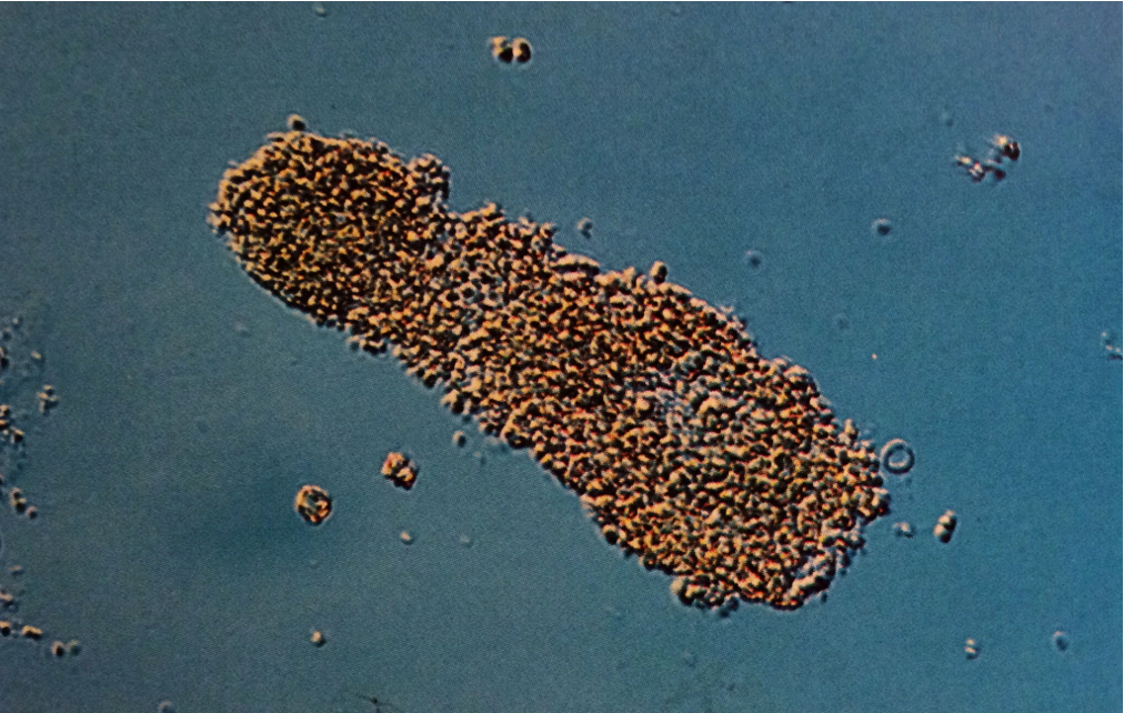
White blood cell cast in which leukocytes are intact and easily identified as having multilobed nuclei. In some areas, hyaline matrix can still be seen and is not obscured by heavy concentration of PMNs
160X
Source: Urinary Sediment: A Textbook Atlas. Haber, Meryl H.

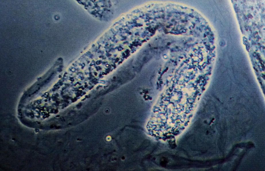
Red blood cell and hemoglobin cast. Erythrocytes have degenerated, resulting in granular, yellowish brown hemoglobin in the cast matrix. However, numerous intact erythrocytes are still present and easily identifiable on the surface
Interference-contrast microscopy, 200X
Source: Urinary Sediment: A Textbook Atlas. Haber, Meryl H.
Red blood cell cast, showing predominately intact erythrocytes closely packed together. Surrounding urine is filled with numerous PMNs and erythrocytes
160X
Source: Urinary Sediment: A Textbook Atlas. Haber, Meryl H.
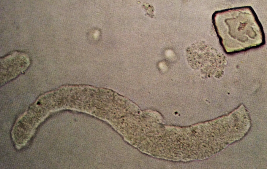
Red blood cell cast. Erythrocytes are more densely concentrated towards one end of hyaline cast matrix
160X
Source: Urinary Sediment: A Textbook Atlas. Haber, Meryl H.
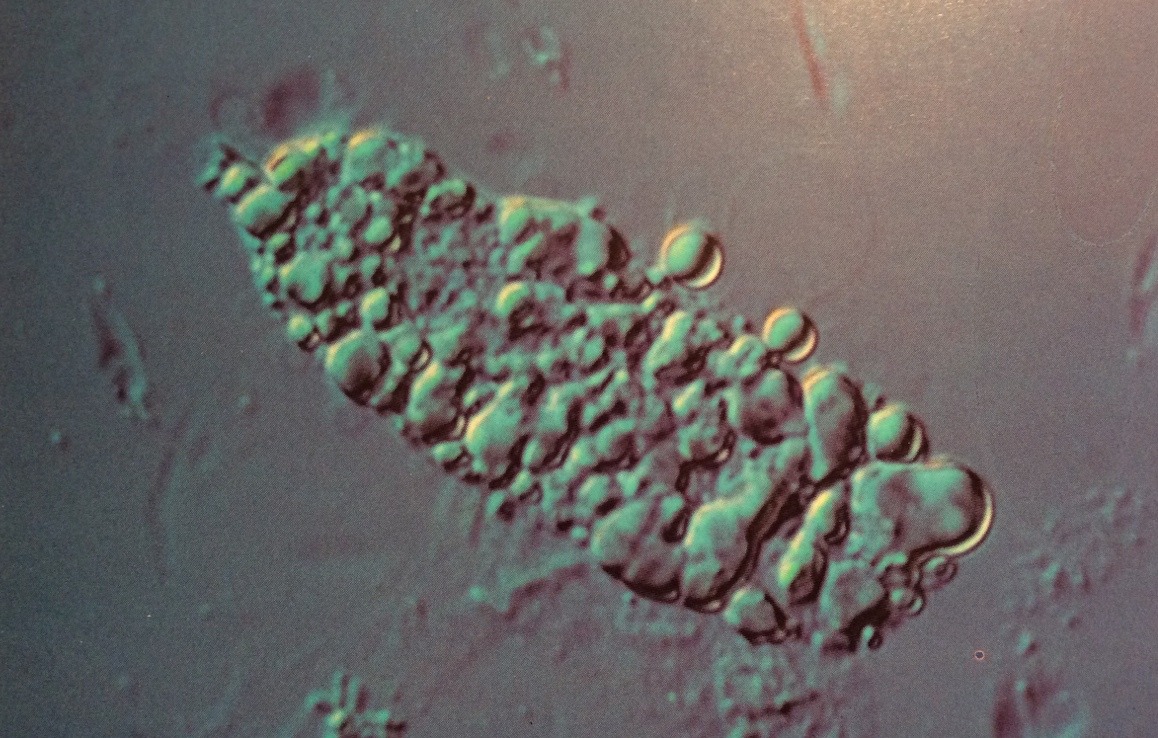
Fatty cast. Lipid globules are prominent on its surface, but some uninvolved surface areas can also be seen underlying the lipids. Some lipid globules are also adjacent to the cast
Interference-contrast microscopy, 400X
Source: Urinary Sediment: A Textbook Atlas. Haber, Meryl H.
Typical fatty cast in urine. Entire surface seems to comprise lipid globules of variable sizes. In the background, are several red cells and considerable cellular debris
200X
Source: Urinary Sediment: A Textbook Atlas. Haber, Meryl H.

Waxy cast, typified by “broken-off” ends and parallel serrated margins. Its brittleness is apparent, as it appears to be “splitting”
Sternheimer-Malbin stian, 160X
Source: Urinary Sediment: A Textbook Atlas. Haber, Meryl H.

Typical waxy cast. Refractive index is high, ends are “broken-off,” and parallel margins are irregular or serrated
250X
Source: Urinary Sediment: A Textbook Atlas. Haber, Meryl H.
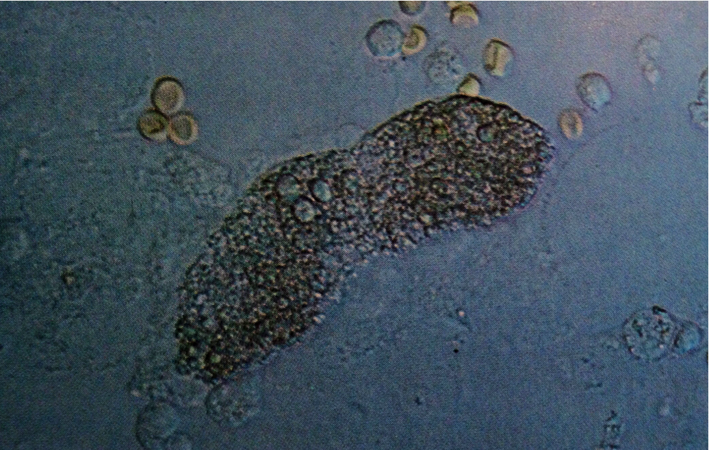
Granular cast revealing certain cellular parts. Note numerous red cells and occasional white blood cells in the background
160X
Source: Urinary Sediment: A Textbook Atlas. Haber, Meryl H.
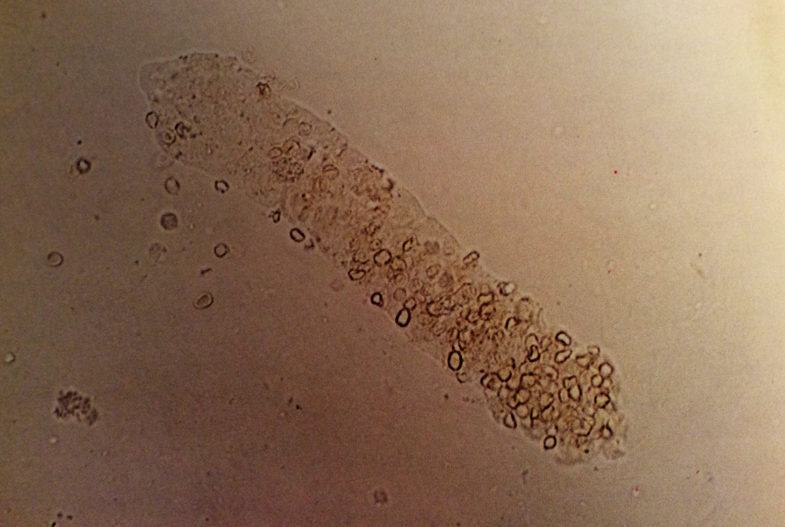
Coarse granular cast, easily seen with interference-contrast microscopy
Interference-contrast microscopy, 160X
Source: Urinary Sediment: A Textbook Atlas. Haber, Meryl H.
Granular cast with numerous mucus threads in the background
Phase-contrast microscopy, 200X
Source: Urinary Sediment: A Textbook Atlas. Haber, Meryl H.

Broad, coarsely granular cast in urine. No vestiges of prior cell origin (i.e., cell cytoplasmic or nuclear components) are seen
Sternheimer-Malbin stain, 200X
Source: Urinary Sediment: A Textbook Atlas. Haber, Meryl H.
Finely granular cast in urine. Also, note uric acid crystal
160X
Source: Urinary Sediment: A Textbook Atlas. Haber, Meryl H.