Quick filters:
Electron Stock Photos and Images
 Colorized scanning electron micrograph of a T lymphocyte. Stock Photohttps://www.alamy.com/image-license-details/?v=1https://www.alamy.com/stock-photo-colorized-scanning-electron-micrograph-of-a-t-lymphocyte-130443046.html
RFHG65C6–Colorized scanning electron micrograph of a T lymphocyte.
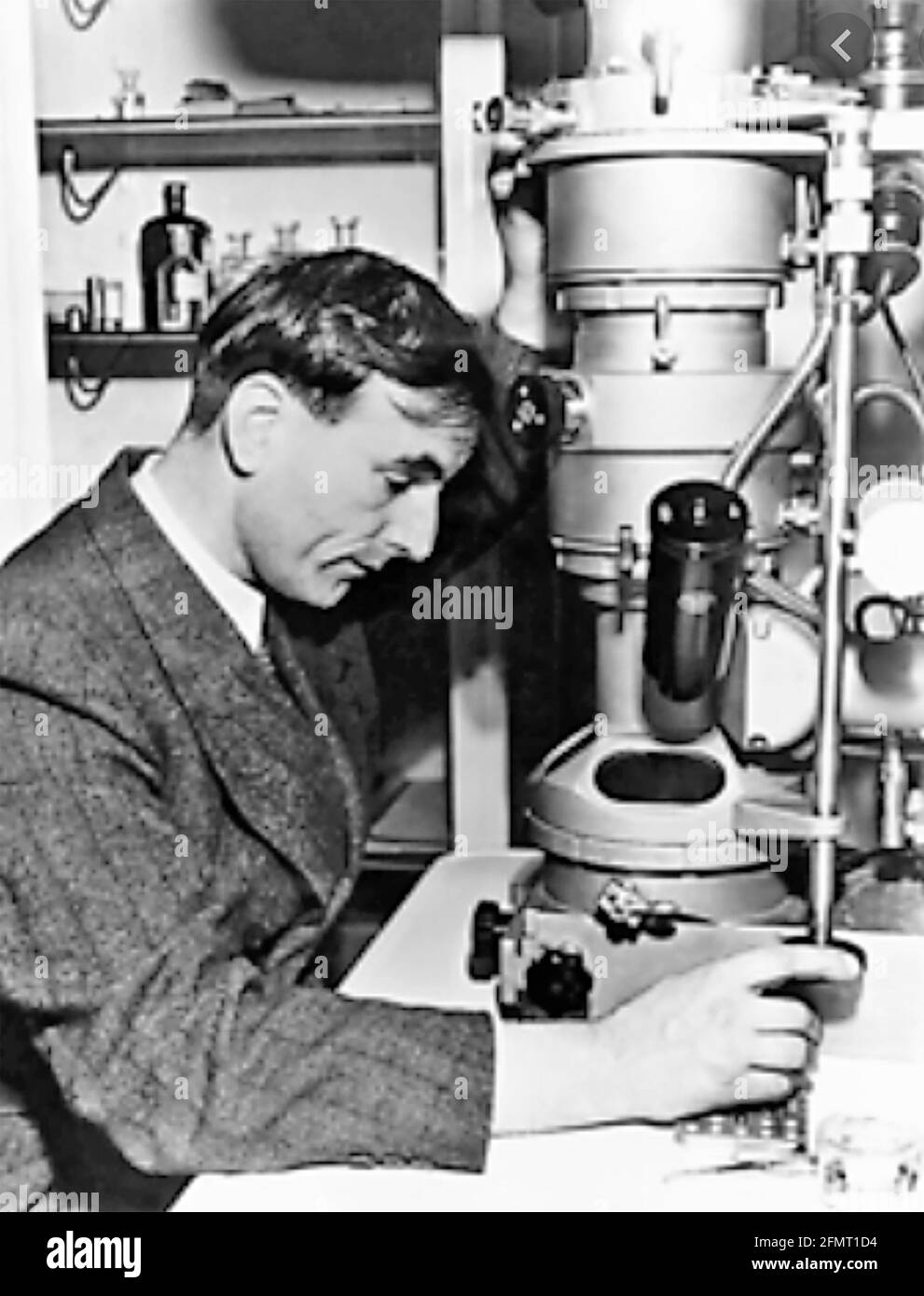 ERNST RUSKA (1906-1988) German physicist who designed the first electron microscope Stock Photohttps://www.alamy.com/image-license-details/?v=1https://www.alamy.com/ernst-ruska-1906-1988-german-physicist-who-designed-the-first-electron-microscope-image425869952.html
RM2FMT1D4–ERNST RUSKA (1906-1988) German physicist who designed the first electron microscope
 Electron Tree or Lichtenberg Figure Stock Photohttps://www.alamy.com/image-license-details/?v=1https://www.alamy.com/electron-tree-or-lichtenberg-figure-image352801652.html
RM2BDYE30–Electron Tree or Lichtenberg Figure
 Nitrogen, atom model. Chemical element with symbol N and with atomic number 7. Bohr model of nitrogen-14. Stock Photohttps://www.alamy.com/image-license-details/?v=1https://www.alamy.com/nitrogen-atom-model-chemical-element-with-symbol-n-and-with-atomic-number-7-bohr-model-of-nitrogen-14-image476287286.html
RF2JJTN86–Nitrogen, atom model. Chemical element with symbol N and with atomic number 7. Bohr model of nitrogen-14.
 atom Stock Photohttps://www.alamy.com/image-license-details/?v=1https://www.alamy.com/stock-photo-atom-14123553.html
RMAG7W1P–atom
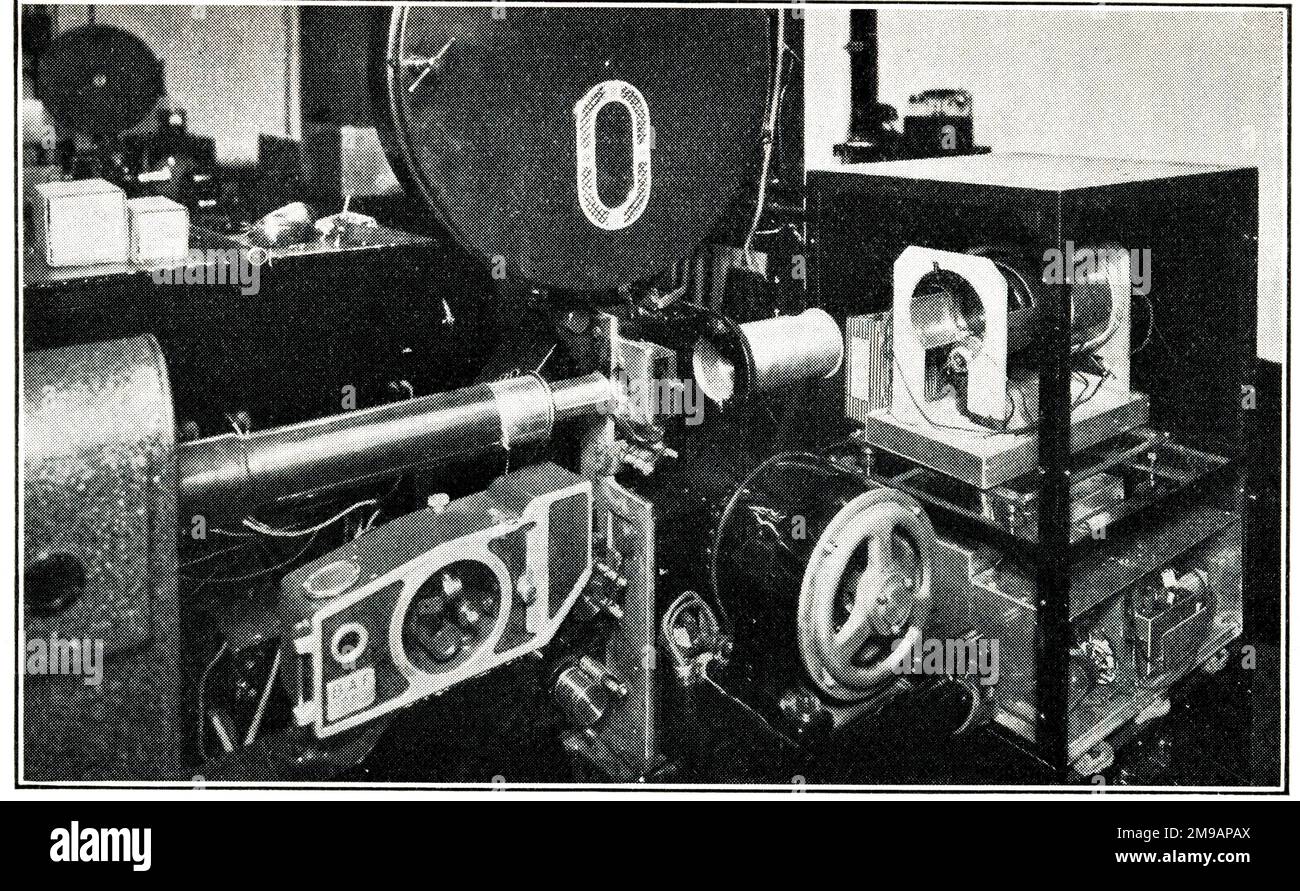 Baird Electron Scanner System of Television, showing the Electron Scanner when used for the televising of talking films. This can be employed for a definition of 100-500 lines. The image dissector for the electron scanning is on the right. There are no mechanically moving parts other than the moving film mechanism. Stock Photohttps://www.alamy.com/image-license-details/?v=1https://www.alamy.com/baird-electron-scanner-system-of-television-showing-the-electron-scanner-when-used-for-the-televising-of-talking-films-this-can-be-employed-for-a-definition-of-100-500-lines-the-image-dissector-for-the-electron-scanning-is-on-the-right-there-are-no-mechanically-moving-parts-other-than-the-moving-film-mechanism-image504869650.html
RM2M9APAX–Baird Electron Scanner System of Television, showing the Electron Scanner when used for the televising of talking films. This can be employed for a definition of 100-500 lines. The image dissector for the electron scanning is on the right. There are no mechanically moving parts other than the moving film mechanism.
 electron microscope in a science lab Stock Photohttps://www.alamy.com/image-license-details/?v=1https://www.alamy.com/stock-photo-electron-microscope-in-a-science-lab-38986245.html
RMC7BY9W–electron microscope in a science lab
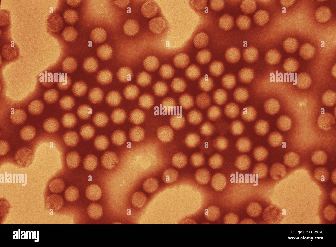 Electron micrograph of equine adenovirus. Stock Photohttps://www.alamy.com/image-license-details/?v=1https://www.alamy.com/stock-photo-electron-micrograph-of-equine-adenovirus-76786634.html
RMECWX3P–Electron micrograph of equine adenovirus.
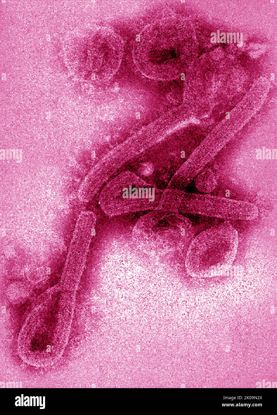 Transmission electron microscopic (TEM) image of Marburg virus virions. Stock Photohttps://www.alamy.com/image-license-details/?v=1https://www.alamy.com/transmission-electron-microscopic-tem-image-of-marburg-virus-virions-image482104418.html
RM2K09N2X–Transmission electron microscopic (TEM) image of Marburg virus virions.
 Broad billed motmot (Electron platyrhynchum platyrhynchum) at feeder with bananas, Mindo cloud forest, Ecuador. Stock Photohttps://www.alamy.com/image-license-details/?v=1https://www.alamy.com/broad-billed-motmot-electron-platyrhynchum-platyrhynchum-at-feeder-with-bananas-mindo-cloud-forest-ecuador-image613563649.html
RF2XJ66KD–Broad billed motmot (Electron platyrhynchum platyrhynchum) at feeder with bananas, Mindo cloud forest, Ecuador.
 Keel-billed Motmot (Electron carinatum) perched on a branch in Costa Rica Stock Photohttps://www.alamy.com/image-license-details/?v=1https://www.alamy.com/stock-image-keel-billed-motmot-electron-carinatum-perched-on-a-branch-in-costa-162651556.html
RFKCHBM4–Keel-billed Motmot (Electron carinatum) perched on a branch in Costa Rica
 Apr. 17, 2012 - Fly's Eye Electron lens: ''Fly's eye'' electron lens that will eventually permit the recording of the contents of three 1,000- page dictionaries in an area the size of a postage stamp is displayed by Sterling P. Newberry, its inventor, at the General Electric Research and Development Center in Scheneotady, New york. The device a compound electron lens two inches in diameter and one-half inch thick, is composed of 1,204 smaller lenses, and is similar in function to a fly's eye (which contains 4,000 small lenses) Stock Photohttps://www.alamy.com/image-license-details/?v=1https://www.alamy.com/apr-17-2012-flys-eye-electron-lens-flys-eye-electron-lens-that-will-image69552091.html
RME14AB7–Apr. 17, 2012 - Fly's Eye Electron lens: ''Fly's eye'' electron lens that will eventually permit the recording of the contents of three 1,000- page dictionaries in an area the size of a postage stamp is displayed by Sterling P. Newberry, its inventor, at the General Electric Research and Development Center in Scheneotady, New york. The device a compound electron lens two inches in diameter and one-half inch thick, is composed of 1,204 smaller lenses, and is similar in function to a fly's eye (which contains 4,000 small lenses)
 Transmission electron Micrograph of the Ebola Virus Hemorrhagic Fever RNA Virus Stock Photohttps://www.alamy.com/image-license-details/?v=1https://www.alamy.com/transmission-electron-micrograph-of-the-ebola-virus-hemorrhagic-fever-rna-virus-image210386207.html
RMP67WN3–Transmission electron Micrograph of the Ebola Virus Hemorrhagic Fever RNA Virus
 Broad-billed Motmot, Electron platyrhynchum, Costa Rica Stock Photohttps://www.alamy.com/image-license-details/?v=1https://www.alamy.com/stock-photo-broad-billed-motmot-electron-platyrhynchum-costa-rica-37909561.html
RMC5JX0W–Broad-billed Motmot, Electron platyrhynchum, Costa Rica
 Transmission electron microscope 'EM9'. Signed: Carl Zeiss. 1964 Stock Photohttps://www.alamy.com/image-license-details/?v=1https://www.alamy.com/stock-photo-transmission-electron-microscope-em9-signed-carl-zeiss-1964-57136848.html
RMD8XPHM–Transmission electron microscope 'EM9'. Signed: Carl Zeiss. 1964
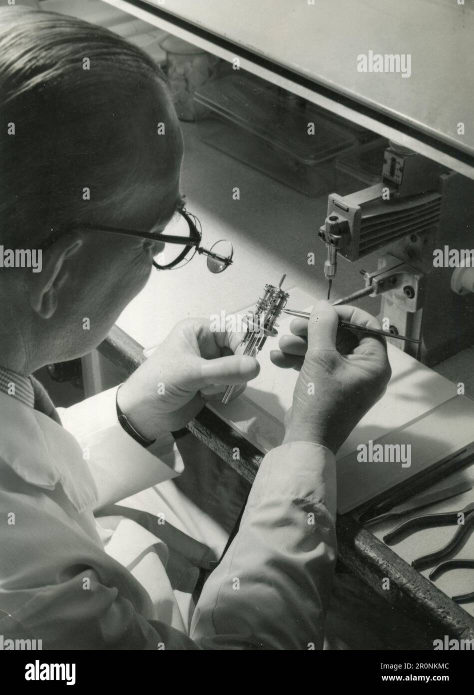 Technician alining the cathode with other electrodes of a travelling wave tube electron gun assembly, UK 1966 Stock Photohttps://www.alamy.com/image-license-details/?v=1https://www.alamy.com/technician-alining-the-cathode-with-other-electrodes-of-a-travelling-wave-tube-electron-gun-assembly-uk-1966-image551208236.html
RF2R0NKMC–Technician alining the cathode with other electrodes of a travelling wave tube electron gun assembly, UK 1966
 A logo sign outside of a facility occupied by Tokyo Electron America, Inc., in Austin, Texas on September 11, 2015. Stock Photohttps://www.alamy.com/image-license-details/?v=1https://www.alamy.com/stock-photo-a-logo-sign-outside-of-a-facility-occupied-by-tokyo-electron-america-87675131.html
RMF2HXEK–A logo sign outside of a facility occupied by Tokyo Electron America, Inc., in Austin, Texas on September 11, 2015.
 Optical electron microscope. Laboratory instrument with clipping path included. Stock Photohttps://www.alamy.com/image-license-details/?v=1https://www.alamy.com/optical-electron-microscope-laboratory-instrument-with-clipping-path-included-image401611874.html
RF2E9B016–Optical electron microscope. Laboratory instrument with clipping path included.
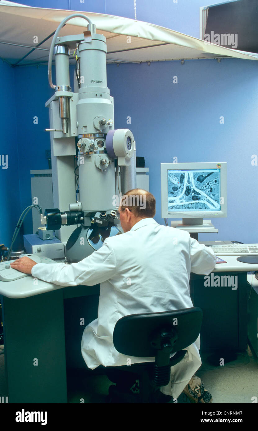 Transmission Electron microscope, scientist/researcher. Stock Photohttps://www.alamy.com/image-license-details/?v=1https://www.alamy.com/stock-photo-transmission-electron-microscope-scientistresearcher-47850439.html
RMCNRNM7–Transmission Electron microscope, scientist/researcher.
 Berlin electron storage ring, Bessy II, Adlershof Science City, Berlin, Germany, Europe Stock Photohttps://www.alamy.com/image-license-details/?v=1https://www.alamy.com/stock-photo-berlin-electron-storage-ring-bessy-ii-adlershof-science-city-berlin-34619507.html
RMC091EY–Berlin electron storage ring, Bessy II, Adlershof Science City, Berlin, Germany, Europe
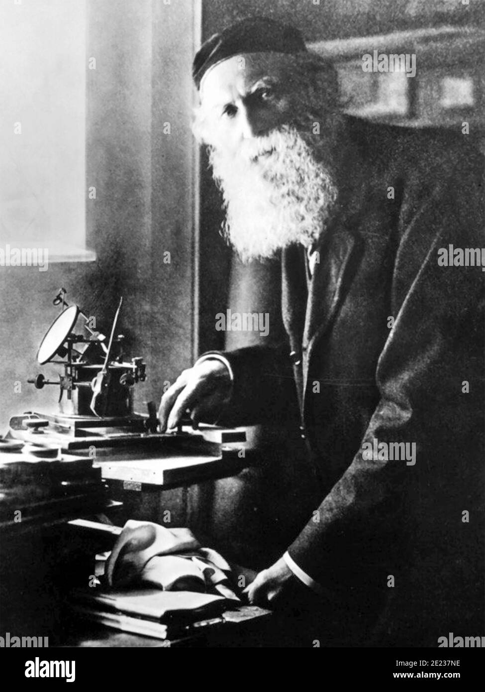 GEORGE JOHNSTONE STONEY (1826-1911) Irish physicist who introduced thew term electron Stock Photohttps://www.alamy.com/image-license-details/?v=1https://www.alamy.com/george-johnstone-stoney-1826-1911-irish-physicist-who-introduced-thew-term-electron-image397139722.html
RM2E237NE–GEORGE JOHNSTONE STONEY (1826-1911) Irish physicist who introduced thew term electron
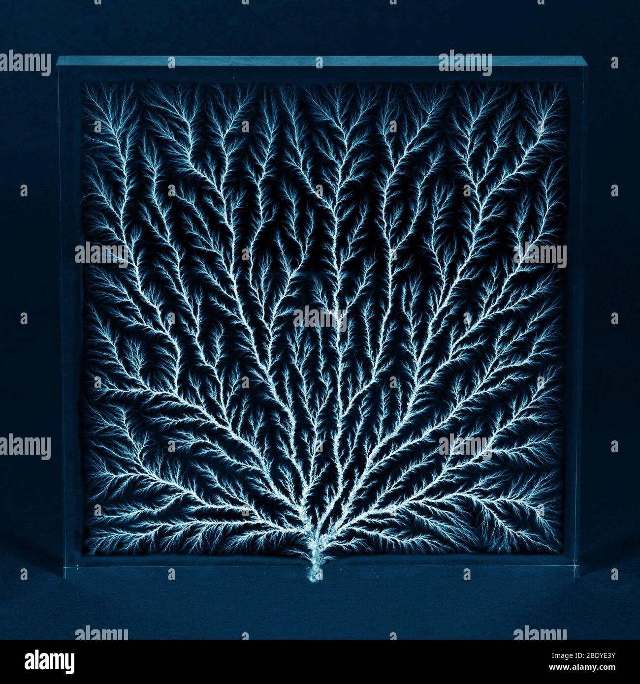 Electron Tree or Lichtenberg Figure Stock Photohttps://www.alamy.com/image-license-details/?v=1https://www.alamy.com/electron-tree-or-lichtenberg-figure-image352801679.html
RM2BDYE3Y–Electron Tree or Lichtenberg Figure
 Clinton Joseph Davisson (1881–1958), was an American physicist who won the 1937 Nobel Prize in Physics (which he shared with George Paget Thomson) for his discovery of electron diffraction in the Davisson-Germer experiment. (Photo: November 1937) Stock Photohttps://www.alamy.com/image-license-details/?v=1https://www.alamy.com/clinton-joseph-davisson-18811958-was-an-american-physicist-who-won-the-1937-nobel-prize-in-physics-which-he-shared-with-george-paget-thomson-for-his-discovery-of-electron-diffraction-in-the-davisson-germer-experiment-photo-november-1937-image210927470.html
RMP74G3X–Clinton Joseph Davisson (1881–1958), was an American physicist who won the 1937 Nobel Prize in Physics (which he shared with George Paget Thomson) for his discovery of electron diffraction in the Davisson-Germer experiment. (Photo: November 1937)
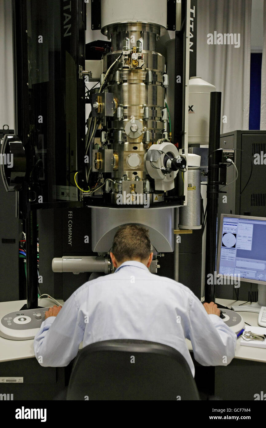 Dr Markus Boese at the controls of a transmission electron microscope as Ireland's most advanced nanoscience research facility, the Crann Advanced Microscopy Laboratory, housing some of the world's most powerful microscopes, officially opens at Trinity Technology and Enterprise Campus in Dublin. Stock Photohttps://www.alamy.com/image-license-details/?v=1https://www.alamy.com/stock-photo-dr-markus-boese-at-the-controls-of-a-transmission-electron-microscope-110973412.html
RMGCF7M4–Dr Markus Boese at the controls of a transmission electron microscope as Ireland's most advanced nanoscience research facility, the Crann Advanced Microscopy Laboratory, housing some of the world's most powerful microscopes, officially opens at Trinity Technology and Enterprise Campus in Dublin.
 Electron micrograph cross section of Escherichia coli bacteria Stock Photohttps://www.alamy.com/image-license-details/?v=1https://www.alamy.com/electron-micrograph-cross-section-of-escherichia-coli-bacteria-image223532950.html
RMPYJPFJ–Electron micrograph cross section of Escherichia coli bacteria
 electron microscope in a science lab Stock Photohttps://www.alamy.com/image-license-details/?v=1https://www.alamy.com/stock-photo-electron-microscope-in-a-science-lab-38986279.html
RMC7BYB3–electron microscope in a science lab
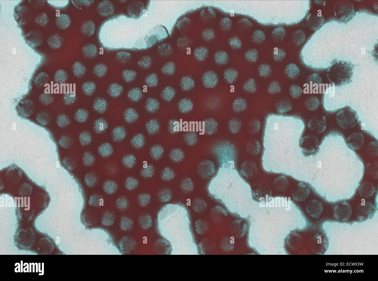 Electron micrograph of equine adenovirus. Stock Photohttps://www.alamy.com/image-license-details/?v=1https://www.alamy.com/stock-photo-electron-micrograph-of-equine-adenovirus-76786637.html
RMECWX3W–Electron micrograph of equine adenovirus.
 Transmission electron Micrograph of the Ebola Virus Hemorrhagic Fever RNA Virus Stock Photohttps://www.alamy.com/image-license-details/?v=1https://www.alamy.com/stock-photo-transmission-electron-micrograph-of-the-ebola-virus-hemorrhagic-fever-76388946.html
RMEC7PTJ–Transmission electron Micrograph of the Ebola Virus Hemorrhagic Fever RNA Virus
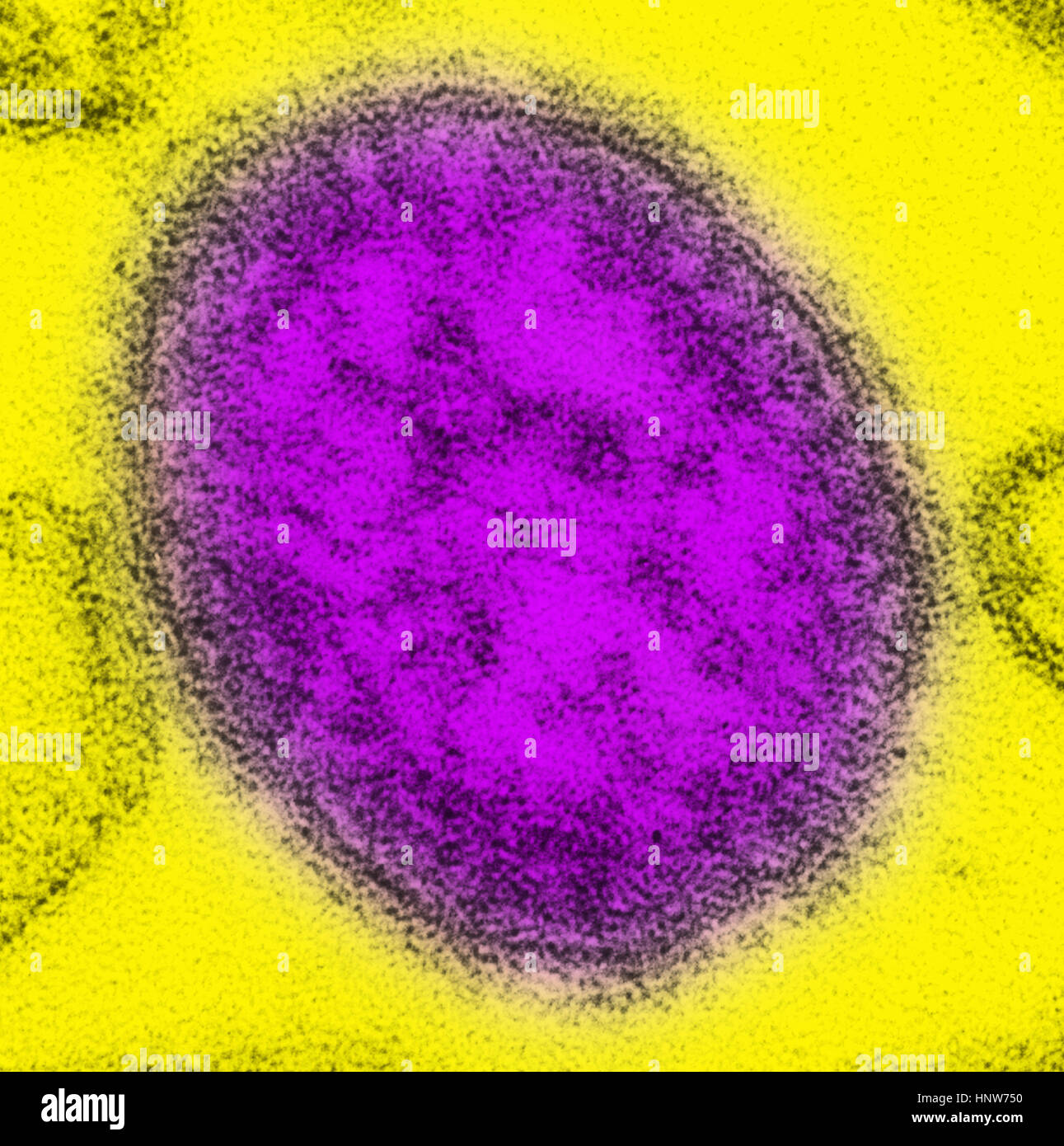 Transmission electron micrograph of a single virus particle, or virion, of measles virus Stock Photohttps://www.alamy.com/image-license-details/?v=1https://www.alamy.com/stock-photo-transmission-electron-micrograph-of-a-single-virus-particle-or-virion-133934780.html
RFHNW750–Transmission electron micrograph of a single virus particle, or virion, of measles virus
 Broad-billed Motmot (Electron platyrhynchum) perched on a branch in Costa Rica Stock Photohttps://www.alamy.com/image-license-details/?v=1https://www.alamy.com/stock-image-broad-billed-motmot-electron-platyrhynchum-perched-on-a-branch-in-162651364.html
RFKCHBD8–Broad-billed Motmot (Electron platyrhynchum) perched on a branch in Costa Rica
 Mar. 22, 2012 - Using this highly advanced electron microscope, Dr. Victor A Phillips right Mr. John A. Hugo, of the General E Stock Photohttps://www.alamy.com/image-license-details/?v=1https://www.alamy.com/mar-22-2012-using-this-highly-advanced-electron-microscope-dr-victor-image69540133.html
RME13R45–Mar. 22, 2012 - Using this highly advanced electron microscope, Dr. Victor A Phillips right Mr. John A. Hugo, of the General E
 Transmission electron Micrograph of the Ebola Virus Hemorrhagic Fever RNA Virus Stock Photohttps://www.alamy.com/image-license-details/?v=1https://www.alamy.com/transmission-electron-micrograph-of-the-ebola-virus-hemorrhagic-fever-rna-virus-image210386206.html
RMP67WN2–Transmission electron Micrograph of the Ebola Virus Hemorrhagic Fever RNA Virus
 Transmission electron micrograph (TEM) of the Sin Nombre virus (SNV), which are members of the genus Hantavirus, within the Stock Photohttps://www.alamy.com/image-license-details/?v=1https://www.alamy.com/stock-photo-transmission-electron-micrograph-tem-of-the-sin-nombre-virus-snv-which-72422131.html
RME5R34K–Transmission electron micrograph (TEM) of the Sin Nombre virus (SNV), which are members of the genus Hantavirus, within the
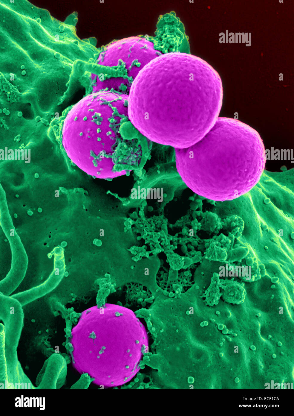 Scanning electron micrograph of a human neutrophil ingesting MRSA. Stock Photohttps://www.alamy.com/image-license-details/?v=1https://www.alamy.com/stock-photo-scanning-electron-micrograph-of-a-human-neutrophil-ingesting-mrsa-76547754.html
RFECF1CA–Scanning electron micrograph of a human neutrophil ingesting MRSA.
 This digitally-colorized transmission electron micrograph (TEM) revealed presence of number of infectious bronchitis virus Stock Photohttps://www.alamy.com/image-license-details/?v=1https://www.alamy.com/stock-photo-this-digitally-colorized-transmission-electron-micrograph-tem-revealed-74194091.html
RME8KR8Y–This digitally-colorized transmission electron micrograph (TEM) revealed presence of number of infectious bronchitis virus
 A logo sign outside of a facility occupied by Tokyo Electron America, Inc., in Austin, Texas on September 11, 2015. Stock Photohttps://www.alamy.com/image-license-details/?v=1https://www.alamy.com/stock-photo-a-logo-sign-outside-of-a-facility-occupied-by-tokyo-electron-america-87675128.html
RMF2HXEG–A logo sign outside of a facility occupied by Tokyo Electron America, Inc., in Austin, Texas on September 11, 2015.
 Optical electron microscope. Laboratory instrument with clipping path included. Stock Photohttps://www.alamy.com/image-license-details/?v=1https://www.alamy.com/optical-electron-microscope-laboratory-instrument-with-clipping-path-included-image401875697.html
RF2E9R0FD–Optical electron microscope. Laboratory instrument with clipping path included.
 classroom Transmission Electron microscope, Tecnai 12 TEM FEI. Stock Photohttps://www.alamy.com/image-license-details/?v=1https://www.alamy.com/stock-photo-classroom-transmission-electron-microscope-tecnai-12-tem-fei-47847260.html
RMCNRHJM–classroom Transmission Electron microscope, Tecnai 12 TEM FEI.
 Radiolarian Coloured scanning electron micrograph (SEM) of the shell of a radiolarian Radiolaria are single-celled protozoans Stock Photohttps://www.alamy.com/image-license-details/?v=1https://www.alamy.com/radiolarian-coloured-scanning-electron-micrograph-sem-of-the-shell-image69881975.html
RFE1KB4R–Radiolarian Coloured scanning electron micrograph (SEM) of the shell of a radiolarian Radiolaria are single-celled protozoans
 GEORGE STONEY (1826-1911) Irish physicist who introduced the term 'electron' Stock Photohttps://www.alamy.com/image-license-details/?v=1https://www.alamy.com/george-stoney-1826-1911-irish-physicist-who-introduced-the-term-electron-image600010486.html
RM2WT4RDX–GEORGE STONEY (1826-1911) Irish physicist who introduced the term 'electron'
 Electron Tree or Lichtenberg Figure Stock Photohttps://www.alamy.com/image-license-details/?v=1https://www.alamy.com/electron-tree-or-lichtenberg-figure-image352801653.html
RM2BDYE31–Electron Tree or Lichtenberg Figure
 Clinton Joseph Davisson (1881–1958), was an American physicist who won the 1937 Nobel Prize in Physics (which he shared with George Paget Thomson) for his discovery of electron diffraction in the Davisson-Germer experiment. (Photo: November 1937) Stock Photohttps://www.alamy.com/image-license-details/?v=1https://www.alamy.com/clinton-joseph-davisson-18811958-was-an-american-physicist-who-won-the-1937-nobel-prize-in-physics-which-he-shared-with-george-paget-thomson-for-his-discovery-of-electron-diffraction-in-the-davisson-germer-experiment-photo-november-1937-image210927472.html
RMP74G40–Clinton Joseph Davisson (1881–1958), was an American physicist who won the 1937 Nobel Prize in Physics (which he shared with George Paget Thomson) for his discovery of electron diffraction in the Davisson-Germer experiment. (Photo: November 1937)
 Periodic Table of Elements, China. The periodic table is a tabular arrangement of the chemical elements, ordered by their atomic number (number of protons in the nucleus), electron configurations, and recurring chemical properties. The table also shows four rectangular blocks: s-, p- d- and f-block. In general, within one row (period) the elements are metals on the left hand side, and non-metals on the right hand side. Stock Photohttps://www.alamy.com/image-license-details/?v=1https://www.alamy.com/periodic-table-of-elements-china-the-periodic-table-is-a-tabular-arrangement-of-the-chemical-elements-ordered-by-their-atomic-number-number-of-protons-in-the-nucleus-electron-configurations-and-recurring-chemical-properties-the-table-also-shows-four-rectangular-blocks-s-p-d-and-f-block-in-general-within-one-row-period-the-elements-are-metals-on-the-left-hand-side-and-non-metals-on-the-right-hand-side-image344274042.html
RM2B0311E–Periodic Table of Elements, China. The periodic table is a tabular arrangement of the chemical elements, ordered by their atomic number (number of protons in the nucleus), electron configurations, and recurring chemical properties. The table also shows four rectangular blocks: s-, p- d- and f-block. In general, within one row (period) the elements are metals on the left hand side, and non-metals on the right hand side.
 Electron Capture Detector (ECD) Machine Stock Photohttps://www.alamy.com/image-license-details/?v=1https://www.alamy.com/electron-capture-detector-ecd-machine-image227767227.html
RMR6FKBR–Electron Capture Detector (ECD) Machine
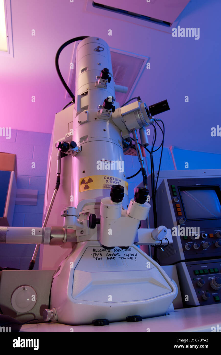 electron microscope in a science lab Stock Photohttps://www.alamy.com/image-license-details/?v=1https://www.alamy.com/stock-photo-electron-microscope-in-a-science-lab-38986250.html
RMC7BYA2–electron microscope in a science lab
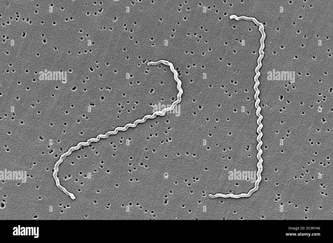 Scanning electron micrograph of Leptospira bacteria. Stock Photohttps://www.alamy.com/image-license-details/?v=1https://www.alamy.com/stock-photo-scanning-electron-micrograph-of-leptospira-bacteria-76787430.html
RMECWY46–Scanning electron micrograph of Leptospira bacteria.
 Transmission electron Micrograph of the Ebola Virus Hemorrhagic Fever RNA Virus Stock Photohttps://www.alamy.com/image-license-details/?v=1https://www.alamy.com/stock-photo-transmission-electron-micrograph-of-the-ebola-virus-hemorrhagic-fever-76388945.html
RMEC7PTH–Transmission electron Micrograph of the Ebola Virus Hemorrhagic Fever RNA Virus
 Broad Billed Motmot (Electron platyrhynchum), Mindo Cloud Forest, Ecuador. Stock Photohttps://www.alamy.com/image-license-details/?v=1https://www.alamy.com/broad-billed-motmot-electron-platyrhynchum-mindo-cloud-forest-ecuador-image553607087.html
RF2R4JYDK–Broad Billed Motmot (Electron platyrhynchum), Mindo Cloud Forest, Ecuador.
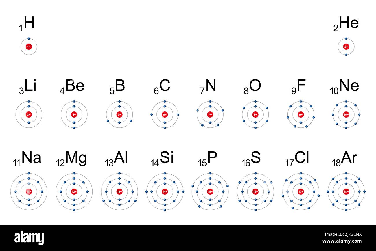 Electron shells of first the 18 chemical elements. An electron shell may be thought of as an orbit followed by electrons around an atomic nucleus. Stock Photohttps://www.alamy.com/image-license-details/?v=1https://www.alamy.com/electron-shells-of-first-the-18-chemical-elements-an-electron-shell-may-be-thought-of-as-an-orbit-followed-by-electrons-around-an-atomic-nucleus-image476434278.html
RF2JK3CNX–Electron shells of first the 18 chemical elements. An electron shell may be thought of as an orbit followed by electrons around an atomic nucleus.
 Apr. 04, 2012 - The JEM Electron microscope is the latest type produced, and can be used for the transmission and reflection of Stock Photohttps://www.alamy.com/image-license-details/?v=1https://www.alamy.com/apr-04-2012-the-jem-electron-microscope-is-the-latest-type-produced-image69546132.html
RME142PC–Apr. 04, 2012 - The JEM Electron microscope is the latest type produced, and can be used for the transmission and reflection of
 Broad-billed Motmot Electron platyrhynchum Tambopata Peru Stock Photohttps://www.alamy.com/image-license-details/?v=1https://www.alamy.com/stock-photo-broad-billed-motmot-electron-platyrhynchum-tambopata-peru-39918721.html
RMC8XCMH–Broad-billed Motmot Electron platyrhynchum Tambopata Peru
 Scanning Electron Micrograph (SEM) of Streptococcus pneumoniae bacteria, This Gram-negative bacterium is a leading cause of Stock Photohttps://www.alamy.com/image-license-details/?v=1https://www.alamy.com/stock-photo-scanning-electron-micrograph-sem-of-streptococcus-pneumoniae-bacteria-72422512.html
RME5R3J8–Scanning Electron Micrograph (SEM) of Streptococcus pneumoniae bacteria, This Gram-negative bacterium is a leading cause of
 Centers for Disease Control and Prevention (CDC) intern, Maureen Metcalfe using transmission electron microscopes (TEM), 2011. Image courtesy Centers for Disease Control / Cynthia Goldsmith. () Stock Photohttps://www.alamy.com/image-license-details/?v=1https://www.alamy.com/centers-for-disease-control-and-prevention-cdc-intern-maureen-metcalfe-using-transmission-electron-microscopes-tem-2011-image-courtesy-centers-for-disease-control-cynthia-goldsmith-image216969118.html
RMPGYP92–Centers for Disease Control and Prevention (CDC) intern, Maureen Metcalfe using transmission electron microscopes (TEM), 2011. Image courtesy Centers for Disease Control / Cynthia Goldsmith. ()
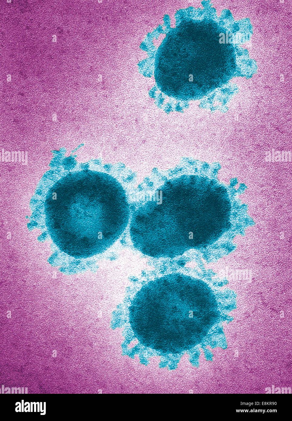 This digitally-colorized transmission electron micrograph (TEM) revealed presence of number of infectious bronchitis virus Stock Photohttps://www.alamy.com/image-license-details/?v=1https://www.alamy.com/stock-photo-this-digitally-colorized-transmission-electron-micrograph-tem-revealed-74194092.html
RME8KR90–This digitally-colorized transmission electron micrograph (TEM) revealed presence of number of infectious bronchitis virus
 A logo sign outside of a facility occupied by Tokyo Electron America, Inc., in Austin, Texas on September 11, 2015. Stock Photohttps://www.alamy.com/image-license-details/?v=1https://www.alamy.com/stock-photo-a-logo-sign-outside-of-a-facility-occupied-by-tokyo-electron-america-87675129.html
RMF2HXEH–A logo sign outside of a facility occupied by Tokyo Electron America, Inc., in Austin, Texas on September 11, 2015.
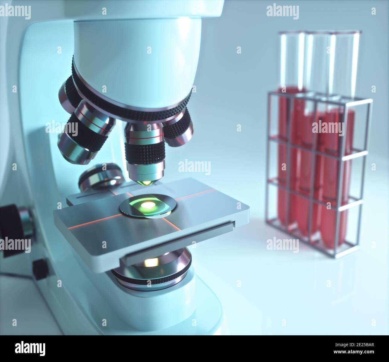 Optical electron microscope. Laboratory instrument, concept of science and microscopic research. Stock Photohttps://www.alamy.com/image-license-details/?v=1https://www.alamy.com/optical-electron-microscope-laboratory-instrument-concept-of-science-and-microscopic-research-image397186463.html
RF2E25BAR–Optical electron microscope. Laboratory instrument, concept of science and microscopic research.
 Broad-billed Motmot (Electron platyrhynchum) Tambopata, Peru Stock Photohttps://www.alamy.com/image-license-details/?v=1https://www.alamy.com/broad-billed-motmot-electron-platyrhynchum-tambopata-peru-image263189980.html
RMW859E4–Broad-billed Motmot (Electron platyrhynchum) Tambopata, Peru
 Coloured scanning electron micrograph (SEM) of Enterococcus faecalis (formerly known as Streptococcus faecalis),Gram positive,coccoid prokaryote (dividing); causes skin wound infections such as scalded skin syndrome,scarlet fever,erysipelas impetigo.Group Streptococcus.Enterococcus faecalis Stock Photohttps://www.alamy.com/image-license-details/?v=1https://www.alamy.com/stock-photo-coloured-scanning-electron-micrograph-sem-of-enterococcus-faecalis-131545318.html
RFHJ0BB2–Coloured scanning electron micrograph (SEM) of Enterococcus faecalis (formerly known as Streptococcus faecalis),Gram positive,coccoid prokaryote (dividing); causes skin wound infections such as scalded skin syndrome,scarlet fever,erysipelas impetigo.Group Streptococcus.Enterococcus faecalis
 JOSEPH JOHN THOMSON (1856-1940) English physicist who discovered the electron, about 1930. Stock Photohttps://www.alamy.com/image-license-details/?v=1https://www.alamy.com/joseph-john-thomson-1856-1940-english-physicist-who-discovered-the-electron-about-1930-image499477209.html
RM2M0H47N–JOSEPH JOHN THOMSON (1856-1940) English physicist who discovered the electron, about 1930.
 Transmission electron micrograph (TEM) of influenza C virus. Stock Photohttps://www.alamy.com/image-license-details/?v=1https://www.alamy.com/transmission-electron-micrograph-tem-of-influenza-c-virus-image352826845.html
RM2BE0J6N–Transmission electron micrograph (TEM) of influenza C virus.
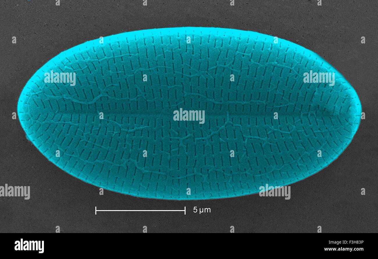 Scanning electron micrograph diatom Stock Photohttps://www.alamy.com/image-license-details/?v=1https://www.alamy.com/stock-photo-scanning-electron-micrograph-diatom-88275370.html
RFF3H83P–Scanning electron micrograph diatom
 Periodic Table of Elements, China. The periodic table is a tabular arrangement of the chemical elements, ordered by their atomic number (number of protons in the nucleus), electron configurations, and recurring chemical properties. The table also shows four rectangular blocks: s-, p- d- and f-block. In general, within one row (period) the elements are metals on the left hand side, and non-metals on the right hand side. Stock Photohttps://www.alamy.com/image-license-details/?v=1https://www.alamy.com/periodic-table-of-elements-china-the-periodic-table-is-a-tabular-arrangement-of-the-chemical-elements-ordered-by-their-atomic-number-number-of-protons-in-the-nucleus-electron-configurations-and-recurring-chemical-properties-the-table-also-shows-four-rectangular-blocks-s-p-d-and-f-block-in-general-within-one-row-period-the-elements-are-metals-on-the-left-hand-side-and-non-metals-on-the-right-hand-side-image344274043.html
RM2B0311F–Periodic Table of Elements, China. The periodic table is a tabular arrangement of the chemical elements, ordered by their atomic number (number of protons in the nucleus), electron configurations, and recurring chemical properties. The table also shows four rectangular blocks: s-, p- d- and f-block. In general, within one row (period) the elements are metals on the left hand side, and non-metals on the right hand side.
 electron storage ring facility BESSY II, Berlin Stock Photohttps://www.alamy.com/image-license-details/?v=1https://www.alamy.com/stock-photo-electron-storage-ring-facility-bessy-ii-berlin-27701727.html
RMBH1WRB–electron storage ring facility BESSY II, Berlin
 electron microscope in a science lab Stock Photohttps://www.alamy.com/image-license-details/?v=1https://www.alamy.com/stock-photo-electron-microscope-in-a-science-lab-38986249.html
RMC7BYA1–electron microscope in a science lab
 Electron micrograph of Human Immunodeficiency Virus, HIV. Stock Photohttps://www.alamy.com/image-license-details/?v=1https://www.alamy.com/stock-photo-electron-micrograph-of-human-immunodeficiency-virus-hiv-76787266.html
RMECWXXA–Electron micrograph of Human Immunodeficiency Virus, HIV.
 transmission electron microscope image Marburg virus virion grown of tissue culture cells Stock Photohttps://www.alamy.com/image-license-details/?v=1https://www.alamy.com/stock-photo-transmission-electron-microscope-image-marburg-virus-virion-grown-76389369.html
RMEC7RBN–transmission electron microscope image Marburg virus virion grown of tissue culture cells
 Broad Billed Motmot (Electron platyrhynchum), Mindo Cloud Forest, Ecuador. Stock Photohttps://www.alamy.com/image-license-details/?v=1https://www.alamy.com/broad-billed-motmot-electron-platyrhynchum-mindo-cloud-forest-ecuador-image553530333.html
RF2R4FDGD–Broad Billed Motmot (Electron platyrhynchum), Mindo Cloud Forest, Ecuador.
 Electron micrograph of a cross section of mitochondria in cardiac muscle tissue Stock Photohttps://www.alamy.com/image-license-details/?v=1https://www.alamy.com/electron-micrograph-of-a-cross-section-of-mitochondria-in-cardiac-muscle-tissue-image223532988.html
RMPYJPH0–Electron micrograph of a cross section of mitochondria in cardiac muscle tissue
 Feb. 26, 2012 - Testing the electron microscopes at the Japan Electron Optics laboratory in Tokyo. The smaller model in the foreground is the JEM T-4. The three others are later JEM-5G. Stock Photohttps://www.alamy.com/image-license-details/?v=1https://www.alamy.com/feb-26-2012-testing-the-electron-microscopes-at-the-japan-electron-image69522158.html
RME13066–Feb. 26, 2012 - Testing the electron microscopes at the Japan Electron Optics laboratory in Tokyo. The smaller model in the foreground is the JEM T-4. The three others are later JEM-5G.
 Broad-billed Motmot Electron platyrhynchum perched on moss covered branch Stock Photohttps://www.alamy.com/image-license-details/?v=1https://www.alamy.com/stock-photo-broad-billed-motmot-electron-platyrhynchum-perched-on-moss-covered-77996777.html
RMEEW1K5–Broad-billed Motmot Electron platyrhynchum perched on moss covered branch
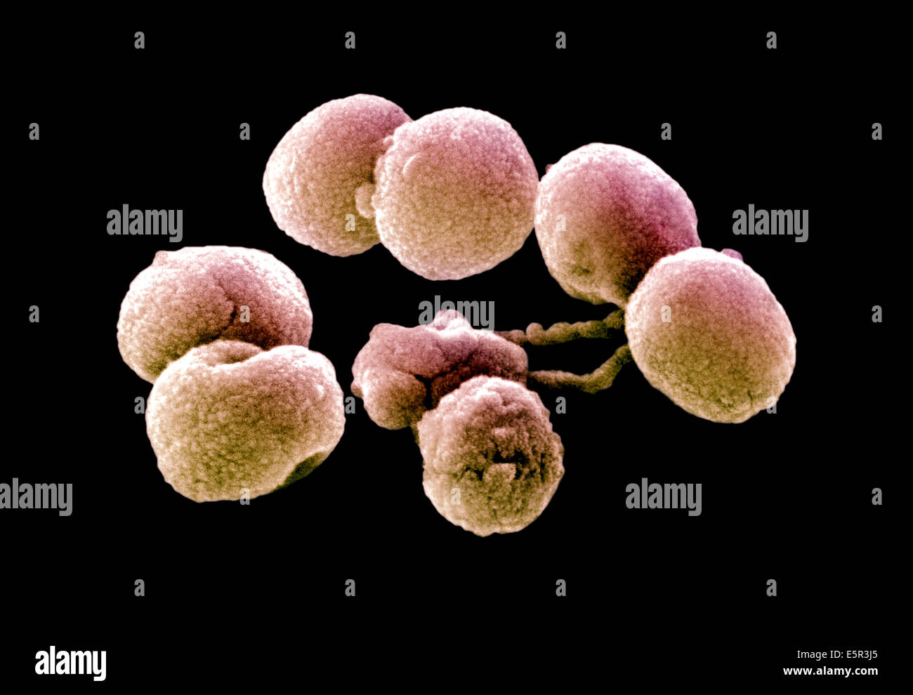 Scanning Electron Micrograph (SEM) of Streptococcus pneumoniae bacteria, This Gram-negative bacterium is a leading cause of Stock Photohttps://www.alamy.com/image-license-details/?v=1https://www.alamy.com/stock-photo-scanning-electron-micrograph-sem-of-streptococcus-pneumoniae-bacteria-72422509.html
RME5R3J5–Scanning Electron Micrograph (SEM) of Streptococcus pneumoniae bacteria, This Gram-negative bacterium is a leading cause of
 Centers for Disease Control and Prevention (CDC) intern, Maureen Metcalfe using transmission electron microscopes (TEM), 2011. Image courtesy Centers for Disease Control / Cynthia Goldsmith. () Stock Photohttps://www.alamy.com/image-license-details/?v=1https://www.alamy.com/centers-for-disease-control-and-prevention-cdc-intern-maureen-metcalfe-using-transmission-electron-microscopes-tem-2011-image-courtesy-centers-for-disease-control-cynthia-goldsmith-image216969115.html
RMPGYP8Y–Centers for Disease Control and Prevention (CDC) intern, Maureen Metcalfe using transmission electron microscopes (TEM), 2011. Image courtesy Centers for Disease Control / Cynthia Goldsmith. ()
 Colorized transmission electron micrograph of Middle East respiratory syndrome coronavirus particles. Stock Photohttps://www.alamy.com/image-license-details/?v=1https://www.alamy.com/stock-photo-colorized-transmission-electron-micrograph-of-middle-east-respiratory-74193859.html
RME8KR0K–Colorized transmission electron micrograph of Middle East respiratory syndrome coronavirus particles.
 A logo sign outside of a facility occupied by Tokyo Electron America, Inc., in Austin, Texas on September 11, 2015. Stock Photohttps://www.alamy.com/image-license-details/?v=1https://www.alamy.com/stock-photo-a-logo-sign-outside-of-a-facility-occupied-by-tokyo-electron-america-87675124.html
RMF2HXEC–A logo sign outside of a facility occupied by Tokyo Electron America, Inc., in Austin, Texas on September 11, 2015.
 Optical electron microscope. Laboratory instrument, concept of science and microscopic research. Stock Photohttps://www.alamy.com/image-license-details/?v=1https://www.alamy.com/optical-electron-microscope-laboratory-instrument-concept-of-science-and-microscopic-research-image397716234.html
RF2E31F36–Optical electron microscope. Laboratory instrument, concept of science and microscopic research.
 A Broad-billed Motmot (Electron platyrhynchum) perched on a branch in Costa Rica Stock Photohttps://www.alamy.com/image-license-details/?v=1https://www.alamy.com/a-broad-billed-motmot-electron-platyrhynchum-perched-on-a-branch-in-costa-rica-image352332520.html
RM2BD63M8–A Broad-billed Motmot (Electron platyrhynchum) perched on a branch in Costa Rica
 Coloured scanning electron micrograph (SEM) of centric fossil diatom frustule.This fresh water diatom frustule came from deposits found at Kalamath Falls,Oregon.This freshwater centric diatom is found in fresh water streams,ponds lakes.Diatoms are type of algae (Chromophyta,Bacillariophyceae).They Stock Photohttps://www.alamy.com/image-license-details/?v=1https://www.alamy.com/stock-photo-coloured-scanning-electron-micrograph-sem-of-centric-fossil-diatom-131545263.html
RFHJ0B93–Coloured scanning electron micrograph (SEM) of centric fossil diatom frustule.This fresh water diatom frustule came from deposits found at Kalamath Falls,Oregon.This freshwater centric diatom is found in fresh water streams,ponds lakes.Diatoms are type of algae (Chromophyta,Bacillariophyceae).They
 BODO von BORRIES (1905-1956) Germany physicist and co-inventor of the electron microscope Stock Photohttps://www.alamy.com/image-license-details/?v=1https://www.alamy.com/bodo-von-borries-1905-1956-germany-physicist-and-co-inventor-of-the-electron-microscope-image425869904.html
RM2FMT1BC–BODO von BORRIES (1905-1956) Germany physicist and co-inventor of the electron microscope
 Transmission electron micrograph (TEM) of influenza A virus. Stock Photohttps://www.alamy.com/image-license-details/?v=1https://www.alamy.com/transmission-electron-micrograph-tem-of-influenza-a-virus-image352826847.html
RM2BE0J6R–Transmission electron micrograph (TEM) of influenza A virus.
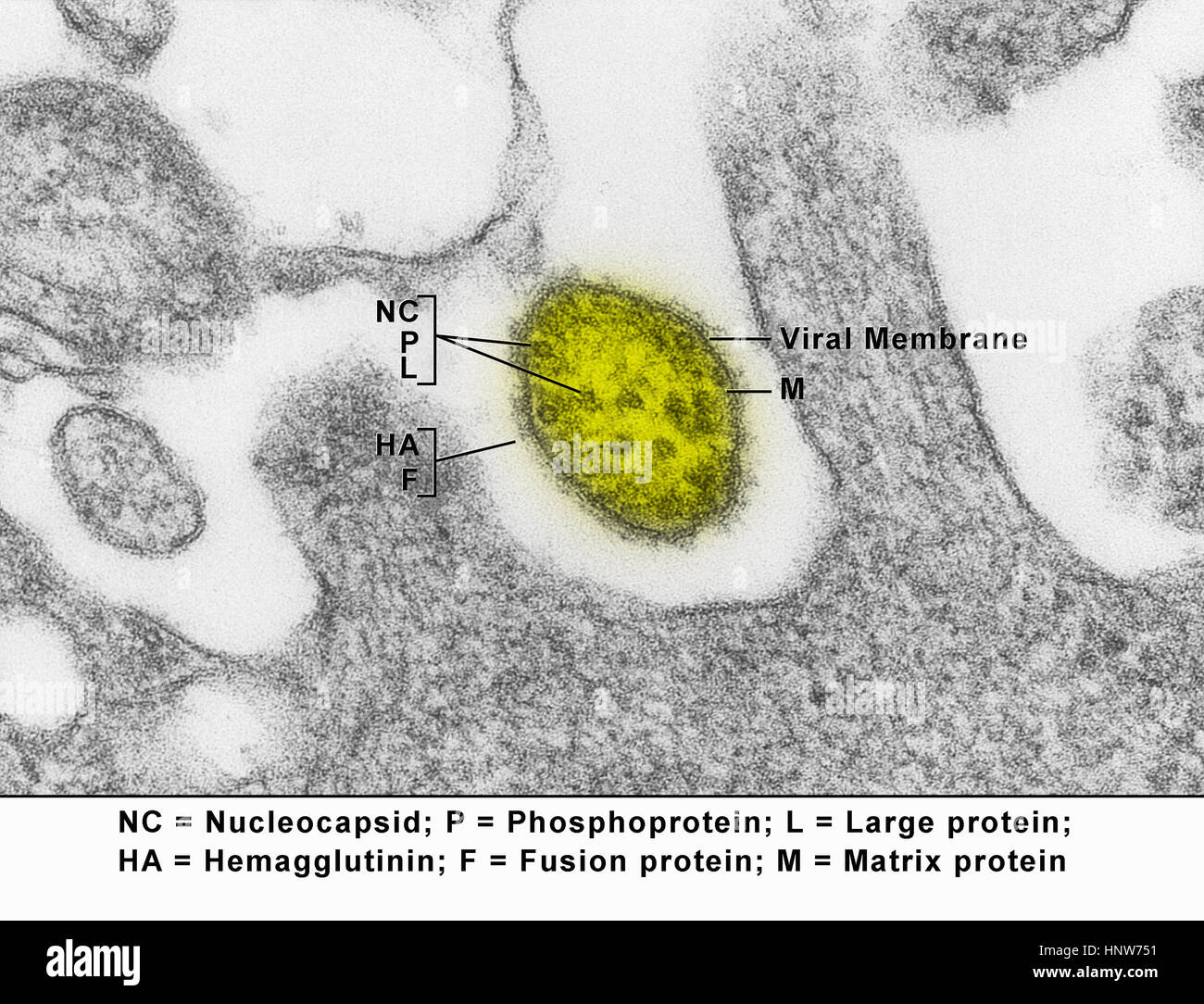 Transmission electron micrograph showing a measles virus particle, or virion Stock Photohttps://www.alamy.com/image-license-details/?v=1https://www.alamy.com/stock-photo-transmission-electron-micrograph-showing-a-measles-virus-particle-133934781.html
RFHNW751–Transmission electron micrograph showing a measles virus particle, or virion
 Plaque commemorationg the discovery of the electron on the Old Cavendish Laboratory, Free School Lane, Cambridge England UK Stock Photohttps://www.alamy.com/image-license-details/?v=1https://www.alamy.com/stock-photo-plaque-commemorationg-the-discovery-of-the-electron-on-the-old-cavendish-23510444.html
RMBA6YPM–Plaque commemorationg the discovery of the electron on the Old Cavendish Laboratory, Free School Lane, Cambridge England UK
 electron storage ring facility BESSY II, Berlin Stock Photohttps://www.alamy.com/image-license-details/?v=1https://www.alamy.com/stock-photo-electron-storage-ring-facility-bessy-ii-berlin-30928028.html
RMBP8W0C–electron storage ring facility BESSY II, Berlin
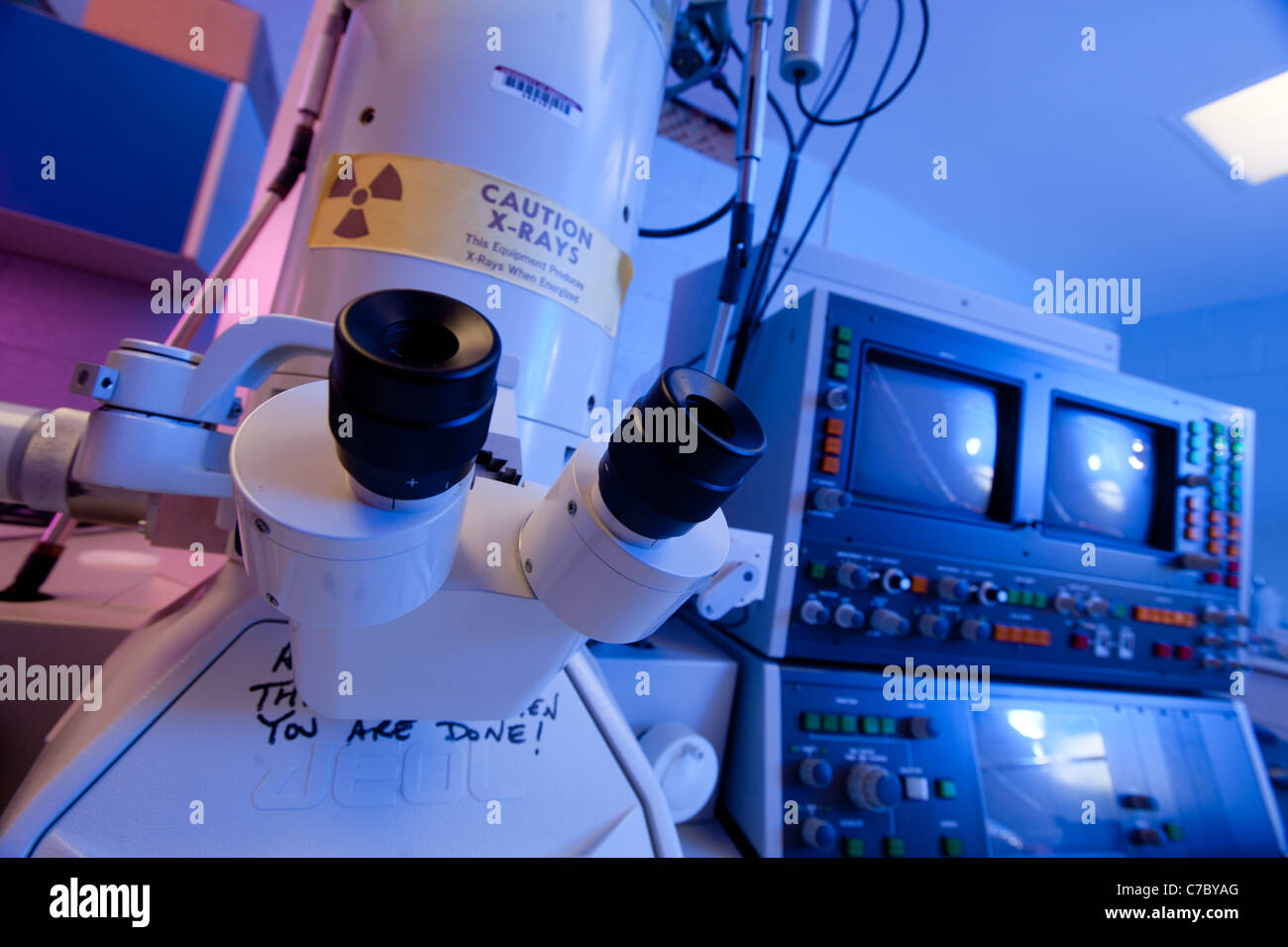 electron microscope in a science lab Stock Photohttps://www.alamy.com/image-license-details/?v=1https://www.alamy.com/stock-photo-electron-microscope-in-a-science-lab-38986264.html
RMC7BYAG–electron microscope in a science lab
 Electron micrograph of Human Immunodeficiency Virus, HIV. Stock Photohttps://www.alamy.com/image-license-details/?v=1https://www.alamy.com/stock-photo-electron-micrograph-of-human-immunodeficiency-virus-hiv-76787260.html
RMECWXX4–Electron micrograph of Human Immunodeficiency Virus, HIV.
 Colorized transmission electron micrograph (TEM), Created by CDC microbiologist Cynthia Goldsmith, ultra structural morphology displayed by an Ebola virus virion Stock Photohttps://www.alamy.com/image-license-details/?v=1https://www.alamy.com/stock-photo-colorized-transmission-electron-micrograph-tem-created-by-cdc-microbiologist-76389676.html
RMEC7RPM–Colorized transmission electron micrograph (TEM), Created by CDC microbiologist Cynthia Goldsmith, ultra structural morphology displayed by an Ebola virus virion
 Broad Billed or Rufous Motmot (Electron platyrhynchum), Mindo Cloud Forest, Ecuador. Stock Photohttps://www.alamy.com/image-license-details/?v=1https://www.alamy.com/broad-billed-or-rufous-motmot-electron-platyrhynchum-mindo-cloud-forest-ecuador-image566001689.html
RF2RTRGX1–Broad Billed or Rufous Motmot (Electron platyrhynchum), Mindo Cloud Forest, Ecuador.
 Old manuals for early basic computers, the Amstrad and Acorn Electron which were part of the home computing market in the 80's Stock Photohttps://www.alamy.com/image-license-details/?v=1https://www.alamy.com/stock-photo-old-manuals-for-early-basic-computers-the-amstrad-and-acorn-electron-78432114.html
RMEFGTXX–Old manuals for early basic computers, the Amstrad and Acorn Electron which were part of the home computing market in the 80's
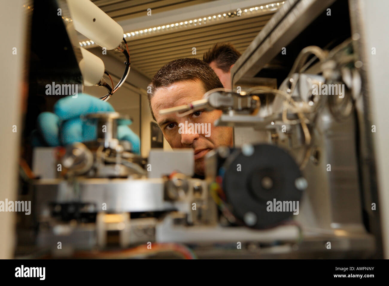 Scientist at the electron microscope IB Cross Bi, Nanostructure Service Laboratory, Karlsruhe university, Baden-Wuerttemberg, G Stock Photohttps://www.alamy.com/image-license-details/?v=1https://www.alamy.com/stock-photo-scientist-at-the-electron-microscope-ib-cross-bi-nanostructure-service-16568534.html
RMAWFNNY–Scientist at the electron microscope IB Cross Bi, Nanostructure Service Laboratory, Karlsruhe university, Baden-Wuerttemberg, G
 Produced by the US National Institute of Allergy and Infectious Diseases (NIAID), this highly magnified, digitally colorized transmission electron microscopic (TEM) image reveals ultrastructural details exhibited by a number of spherical shaped, Middle East respiratory syndrome coronavirus (MERS-CoV) virions. An optimised and enhanced version of an image produced by the US National Institute of Allergy and Infectious Diseases / Credit: NIAID Stock Photohttps://www.alamy.com/image-license-details/?v=1https://www.alamy.com/produced-by-the-us-national-institute-of-allergy-and-infectious-diseases-niaid-this-highly-magnified-digitally-colorized-transmission-electron-microscopic-tem-image-reveals-ultrastructural-details-exhibited-by-a-number-of-spherical-shaped-middle-east-respiratory-syndrome-coronavirus-mers-cov-virions-an-optimised-and-enhanced-version-of-an-image-produced-by-the-us-national-institute-of-allergy-and-infectious-diseases-credit-niaid-image434804493.html
RM2G7B1FW–Produced by the US National Institute of Allergy and Infectious Diseases (NIAID), this highly magnified, digitally colorized transmission electron microscopic (TEM) image reveals ultrastructural details exhibited by a number of spherical shaped, Middle East respiratory syndrome coronavirus (MERS-CoV) virions. An optimised and enhanced version of an image produced by the US National Institute of Allergy and Infectious Diseases / Credit: NIAID
 Coloured scanning electron micrograph (SEM) of methicillin-resistant Staphylococcus aureus (MRSA) bacteria. Stock Photohttps://www.alamy.com/image-license-details/?v=1https://www.alamy.com/stock-photo-coloured-scanning-electron-micrograph-sem-of-methicillin-resistant-79757149.html
RMEHN71H–Coloured scanning electron micrograph (SEM) of methicillin-resistant Staphylococcus aureus (MRSA) bacteria.
 Centers for Disease Control and Prevention (CDC) intern, Maureen Metcalfe using transmission electron microscopes (TEM), 2011. Image courtesy Centers for Disease Control / Cynthia Goldsmith. () Stock Photohttps://www.alamy.com/image-license-details/?v=1https://www.alamy.com/centers-for-disease-control-and-prevention-cdc-intern-maureen-metcalfe-using-transmission-electron-microscopes-tem-2011-image-courtesy-centers-for-disease-control-cynthia-goldsmith-image216969110.html
RMPGYP8P–Centers for Disease Control and Prevention (CDC) intern, Maureen Metcalfe using transmission electron microscopes (TEM), 2011. Image courtesy Centers for Disease Control / Cynthia Goldsmith. ()
 Colorized transmission electron micrograph showing particles of Middle East respiratory syndrome coronavirus that emerged in Stock Photohttps://www.alamy.com/image-license-details/?v=1https://www.alamy.com/stock-photo-colorized-transmission-electron-micrograph-showing-particles-of-middle-74193869.html
RME8KR11–Colorized transmission electron micrograph showing particles of Middle East respiratory syndrome coronavirus that emerged in
 A logo sign outside of a facility occupied by Tokyo Electron America, Inc., in Austin, Texas on September 11, 2015. Stock Photohttps://www.alamy.com/image-license-details/?v=1https://www.alamy.com/stock-photo-a-logo-sign-outside-of-a-facility-occupied-by-tokyo-electron-america-87675127.html
RMF2HXEF–A logo sign outside of a facility occupied by Tokyo Electron America, Inc., in Austin, Texas on September 11, 2015.
 Optical electron microscope. Laboratory instrument, concept of science and microscopic research. Stock Photohttps://www.alamy.com/image-license-details/?v=1https://www.alamy.com/optical-electron-microscope-laboratory-instrument-concept-of-science-and-microscopic-research-image397206731.html
RF2E2696K–Optical electron microscope. Laboratory instrument, concept of science and microscopic research.
 lithium atom colorcode atomic nucleus red=proton, white=neutron, electron shell blue=electron Stock Photohttps://www.alamy.com/image-license-details/?v=1https://www.alamy.com/stock-photo-lithium-atom-colorcode-atomic-nucleus-red=proton-white=neutron-electron-29432657.html
RMBKTNJ9–lithium atom colorcode atomic nucleus red=proton, white=neutron, electron shell blue=electron
 Coloured scanning electron micrograph (SEM) of centric fossil diatom frustule.This fresh water diatom frustule came from deposits found at Kalamath Falls,Oregon.This freshwater centric diatom is found in fresh water streams,ponds lakes.Diatoms are type of algae (Chromophyta,Bacillariophyceae).They Stock Photohttps://www.alamy.com/image-license-details/?v=1https://www.alamy.com/stock-photo-coloured-scanning-electron-micrograph-sem-of-centric-fossil-diatom-131545262.html
RFHJ0B92–Coloured scanning electron micrograph (SEM) of centric fossil diatom frustule.This fresh water diatom frustule came from deposits found at Kalamath Falls,Oregon.This freshwater centric diatom is found in fresh water streams,ponds lakes.Diatoms are type of algae (Chromophyta,Bacillariophyceae).They
 ERNST RUSKA (1906-1988) German physicist who designed the first electron microscope Stock Photohttps://www.alamy.com/image-license-details/?v=1https://www.alamy.com/ernst-ruska-1906-1988-german-physicist-who-designed-the-first-electron-microscope-image425869955.html
RM2FMT1D7–ERNST RUSKA (1906-1988) German physicist who designed the first electron microscope
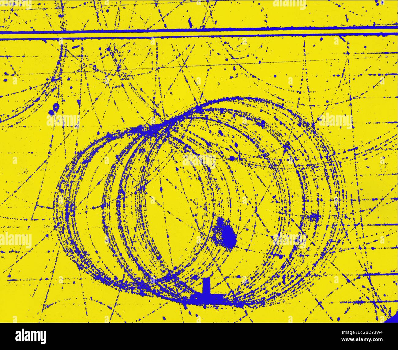 Cloud Chamber Event, Electron and Positron Stock Photohttps://www.alamy.com/image-license-details/?v=1https://www.alamy.com/cloud-chamber-event-electron-and-positron-image352793648.html
RM2BDY3W4–Cloud Chamber Event, Electron and Positron
 A Broad-billed Motmot (Electron platyrhynchum) perched on a branch. Costa Rica. Stock Photohttps://www.alamy.com/image-license-details/?v=1https://www.alamy.com/a-broad-billed-motmot-electron-platyrhynchum-perched-on-a-branch-costa-rica-image455040474.html
RF2HC8TNE–A Broad-billed Motmot (Electron platyrhynchum) perched on a branch. Costa Rica.
