SlideShare a Scribd company logo
By :- Dr. Bindesh D. Patel, PT
Deputy Registrar
P P Savani University
Inter-trochanteric fracture
• May involve either greater trochanter or lesser
trochanter or both
• Elderly by minor force
• Adult by major force
• Femoral neck shaft
angle is reduced (Coxa vara)
• Comminuted and displaced fracture
• Very rarely undisplaced fracture
Diagnosis
• Clinical features
• History of fall or accident
• Pain, swelling, tenderness
• Inability to move leg
• Physical findings are more marked compared to
neck of femur fracture
• Easy to diagnose on X-ray
• Comminution of the medial
cortex of the neck of femur
• Avulsion of lesser
trochanter
• Extension of the fracture of
the sub trochanteric region
indicates unstable fracture
and poor prognosis
Treatment
• It unites readily
• Main objective is to maintain a normal
femoral neck shaft angle
• In elderly patient, ORIF is preferred to prevent
complications
• Conservative method
– Russell’s traction and
– Thomas splint
• Operative method
– Internal fixation used are
• Dynamic Hip Screw
• Ender’s nail
• Gamma nail
– External fixation is used for patients with bed
sores
12. Inter trochanteric fracture
Complications
• Malunion
– Coxa vara
– Elderly mild limping while walking and shortening
– Suitable shoe raise
– Adult
– Corrective osteotomy
• Osteoarthritis
– Due to change in hip biomechanics
– Pain and stiffness
– Physiotherapy treatment
– Later trochanteric osteotomy or THR
Thank you
Please share the video and subscribe my
channel.

More Related Content

12. Inter trochanteric fracture

  • 1. By :- Dr. Bindesh D. Patel, PT Deputy Registrar P P Savani University Inter-trochanteric fracture
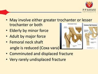
  • 2. • May involve either greater trochanter or lesser trochanter or both • Elderly by minor force • Adult by major force • Femoral neck shaft angle is reduced (Coxa vara) • Comminuted and displaced fracture • Very rarely undisplaced fracture
  • 3. Diagnosis • Clinical features • History of fall or accident • Pain, swelling, tenderness • Inability to move leg • Physical findings are more marked compared to neck of femur fracture
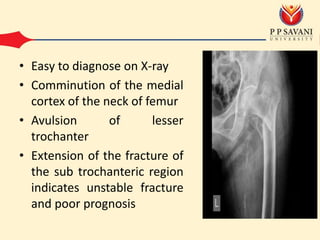
  • 4. • Easy to diagnose on X-ray • Comminution of the medial cortex of the neck of femur • Avulsion of lesser trochanter • Extension of the fracture of the sub trochanteric region indicates unstable fracture and poor prognosis
  • 5. Treatment • It unites readily • Main objective is to maintain a normal femoral neck shaft angle • In elderly patient, ORIF is preferred to prevent complications
  • 6. • Conservative method – Russell’s traction and – Thomas splint
  • 7. • Operative method – Internal fixation used are • Dynamic Hip Screw • Ender’s nail • Gamma nail – External fixation is used for patients with bed sores
  • 10. Complications • Malunion – Coxa vara – Elderly mild limping while walking and shortening – Suitable shoe raise – Adult – Corrective osteotomy
  • 11. • Osteoarthritis – Due to change in hip biomechanics – Pain and stiffness – Physiotherapy treatment – Later trochanteric osteotomy or THR
  • 13. Please share the video and subscribe my channel.