Cerebellum.ppt
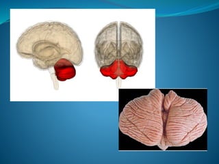
- 2. Latin for ‘little brain’ Largest part of hindbrain Occupies most of posterior cranial fossa Lies behind pons & medulla forming roof of 4th ventricle Separated from posterior part of cerebrum by tentorium cerebelli
- 4. Connection with the brainstem Superior Cerebellar Peduncle (brachium conjunctivum) Connects with midbrain Middle Cerebellar Peduncle (brachium pontis) Connects with pons Inferior Cerebellar Peduncle (restiform body) Connects with medulla
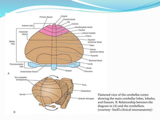
- 5. Two cerebellar hemispheres . Median vermis. Two surfaces ----superior and inferior 3 fissures: fissura prima, horizontal fissure and posterolateral fissure 3 lobes in each hemisphere anterior , posterior and flocculonodular.
- 6. Flattened view of the cerebellar cortex showing the main cerebellar lobes, lobules, and fissures. B: Relationship between the diagram in (A) and the cerebellum. (courtesy- Snell’s clinical neuroanatomy)
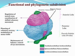
- 7. Functional and phylogenetic subdivision Regulation of muscle tone, coordination of skilled voluntary movement Planning of voluntary activity/ modulation of motor skills Maintenance of balance, control of eye movements Vestibulocerebellum/Archicerebellum Spinocerebellum/Paleocerebellum Cerebrocerebelum/neocerebellum Anterior Lobe Posterior Lobe Flocculo-Nodular Lobe (FN lobe) Primary fissure
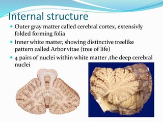
- 8. Internal structure Outer gray matter called cerebral cortex, extensivly folded forming folia Inner white matter, showing distinctive treelike pattern called Arbor vitae (tree of life) 4 pairs of nuclei within white matter ,the deep cerebral nuclei
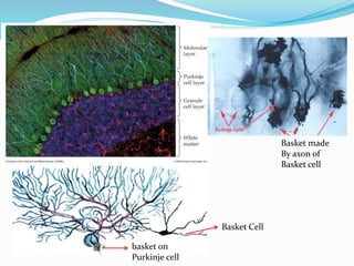
- 9. The cerebral cortex Microscopically composed of three layers: Outer molecular Middle ,Purkinje cell layer and Inner , Granule cell layer Contains five types of neurons: Basket cell Stellate cells Pukinje cell – Purkinje cell layer Granule cell & Golgi cell Purkinje cell axons, are the only output from the cerebellar cortex, to the deep nuclei Molecular layer Granule cell layer
- 10. Basket made By axon of Basket cell Basket Cell basket on Purkinje cell
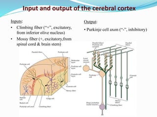
- 11. Inputs: • Climbing fiber (“+”, excitatory, from inferior olive nucleus) • Mossy fiber (+, excitatory,from spinal cord & brain stem) Output: • Purkinje cell axon (“-”, inhibitory) Input and output of the cerebral cortex
- 12. The Afferent Cerebellar Pathways Afferent Tracts Transmits Destination Dorsal spinocerebellar Unconscious kinesthetic & cutaneous afferents from trunk & leg Anterior lobe,pyramis,uvula & paramedian lobe via ipsilateral inferior cerebral peduncle Ventral Spinocerebellar Exteroceptive and proprioceptive fibres from body Vermis and anterior lobe via ipsilateral superior cerebral peduncle Vestibulocerebellar tract Vestibular impulse from labyrinth direct and via vestibular nuclei Flocculonodular lobe via ipsilateral inferior cerebral peduncle Cuneocerebellar tract Proprioceptive impulses, especially from arm,head and neck anterior lobe via ipsilateral inferior cerebral peduncle Tectocerebellar Auditory and visual impulses via inferior and superior colliculi Lobulus simplex ,declive & tuber via superior cerebral peduncle Corticopontocerebellar Impulses from motor and other parts of cerebral cortex via pontine nuclei All parts of cerebellar cortex except flocculonodular lobe via contralateral middle cerebellar peduncle Olivocerebellar Proprioceptive input from whole body via relay in inferior olive All parts of cerebellar cortex via contralateral inferior cerebral peduncle
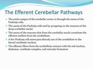
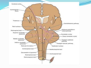
- 13. The Efferent Cerebellar Pathways The entire output of the cerebellar cortex is through the axons of the Purkinje cells. The axons of the Purkinje cells end by synapsing on the neurons of the deep cerebellar nuclei. The axons of the neurons that form the cerebellar nuclei constitute the efferent outflow from the cerebellum. A few Purkinje cell axons pass directly out of the cerebellum to the lateral vestibular nucleus. The efferent fibers from the cerebellum connect with the red nucleus, thalamus, vestibular complex, and reticular formation
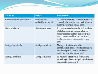
- 14. The Efferent Cerebellar PathwaysPathway Origin Destination Globose-emboliform-rubral Globose and emboliform nuclei To contralateral red nucleus, then via crossed rubrospinal tract to ipsilateral motor neurons in spinal cord Dentothalamic Dentate nucleus To contralateral ventrolateral nucleus of thalamus, then to contralateral motor cerebral cortex; corticospinal tract crosses midline and controls ipsilateral motor neurons in spinal cord Fastigial vestibular Fastigial nucleus Mainly to ipsilateral and to contralateral lateral vestibular nuclei; vestibulospinal tract to ipsilateral motor neurons in spinal cord Fastigial reticular Fastigial nucleus To neurons of reticular formation; reticulospinal tract to ipsilateral motor neurons to spinal cord
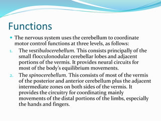
- 17. Functions The nervous system uses the cerebellum to coordinate motor control functions at three levels, as follows: 1. The vestibulocerebellum. This consists principally of the small flocculonodular cerebellar lobes and adjacent portions of the vermis. It provides neural circuits for most of the body’s equilibrium movements. 2. The spinocerebellum. This consists of most of the vermis of the posterior and anterior cerebellum plus the adjacent intermediate zones on both sides of the vermis. It provides the circuitry for coordinating mainly movements of the distal portions of the limbs, especially the hands and fingers.
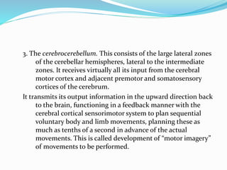
- 18. 3. The cerebrocerebellum. This consists of the large lateral zones of the cerebellar hemispheres, lateral to the intermediate zones. It receives virtually all its input from the cerebral motor cortex and adjacent premotor and somatosensory cortices of the cerebrum. It transmits its output information in the upward direction back to the brain, functioning in a feedback manner with the cerebral cortical sensorimotor system to plan sequential voluntary body and limb movements, planning these as much as tenths of a second in advance of the actual movements. This is called development of “motor imagery” of movements to be performed.
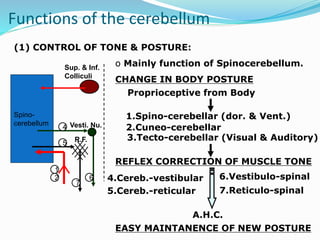
- 19. (1) CONTROL OF TONE & POSTURE: Spino- cerebellum Sup. & Inf. Colliculi Vesti. Nu. R.F. 1 2 4 5 6 7 o Mainly function of Spinocerebellum. CHANGE IN BODY POSTURE Proprioceptive from Body 2.Cuneo-cerebellar 1.Spino-cerebellar (dor. & Vent.) REFLEX CORRECTION OF MUSCLE TONE EASY MAINTANENCE OF NEW POSTURE 3.Tecto-cerebellar (Visual & Auditory) 4.Cereb.-vestibular 5.Cereb.-reticular 6.Vestibulo-spinal 7.Reticulo-spinal A.H.C. Functions of the cerebellum
- 20. (2) CONTROL OF EQUILIBRIUM: o Mainly function of Vestibulocerebellum. Vestibulo- cerebellum Vestibular Apparatus V. N. 1 1 2 3 CHANGE IN HEAD POSITION / ACCELARATION Labyrinthine Afferents 1.Vestibulo-cerebellar REFLEX CORRECTION OF MUSCLE TONE A.H.C. MAINTANENCE OF BODY EQUILIBRIUM 2.Cereb.-vestibular 3.Vestibulo-spinal Function of the cerebellum
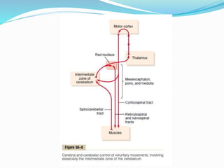
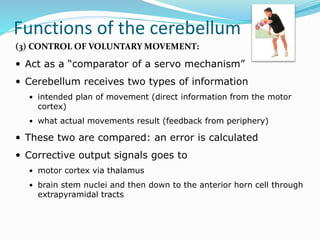
- 21. Functions of the cerebellum (3) CONTROL OF VOLUNTARY MOVEMENT: Act as a “comparator of a servo mechanism” Cerebellum receives two types of information intended plan of movement (direct information from the motor cortex) what actual movements result (feedback from periphery) These two are compared: an error is calculated Corrective output signals goes to motor cortex via thalamus brain stem nuclei and then down to the anterior horn cell through extrapyramidal tracts
- 22. Functions of the cerebellum Corrective Signals through Dentato- Thalamo-Cortical Fibres Cortico-Ponto-Cerebellar fibres Spino-Cerebellar Fibre.
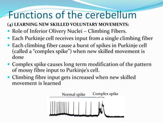
- 23. Functions of the cerebellum (4) LEARNING NEW SKILLED VOLUNTARY MOVEMENTS: Role of Inferior Olivery Nuclei – Climbing Fibers. Each Purkinje cell receives input from a single climbing fiber Each climbing fiber cause a burst of spikes in Purkinje cell (called a “complex spike”) when new skilled movement is done Complex spike causes long term modification of the pattern of mossy fibre input to Purkinje’s cell. Climbing fibre input gets increased when new skilled movement is learned Normal spike Complex spike
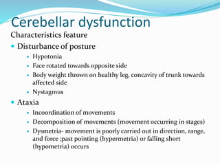
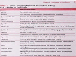
- 24. Cerebellar dysfunction Characteristics feature Disturbance of posture Hypotonia Face rotated towards opposite side Body weight thrown on healthy leg, concavity of trunk towards affected side Nystagmus Ataxia Incoordination of movements Decomposition of movements (movement occurring in stages) Dysmetria- movement is poorly carried out in direction, range, and force ;past pointing (hypermetria) or falling short (hypometria) occurs
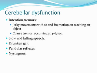
- 25. Cerebellar dysfunction Intention tremors: Jerky movements with to and fro motion on reaching an object Coarse tremor occurring at 4-6/sec. Slow and lalling speech. Drunken gait Pendular reflexes Nystagmus
- 27. Physiotherapy management Head and trunk control Sitting balance Supine to prone Sitting to standing Standing balance/ambulation Ataxia rehabilitation Increase proprioception Increase balance Vestibular exercises
