Tension pneumothorax
- 1. Presented by : BHAVIKA SAHU Roll No. : 25 MBBS FINAL YEAR II DEPARTMENT OF SURGERY Lt.BRKM GMC JDP
- 2. Introduction Incidence Etiology Pathophysiology Clinical features -symptoms -signs Management Complications Differential diagnosis Bibliography
- 3. Pneumothorax is the presence of air between the layers of pleura. It is the most common cause of respiratory insufficiency following chest trauma. Pneumothorax can be – - closed(or simple) - open - tension
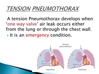
- 5. A tension Pneumothorax develops when ‘one way valve’ air leak occurs either from the lung or through the chest wall. It is an emergency condition.
- 6. Patients with trauma tend to have an associated pneumothorax or tension pneumothorax 20% of the time. In cases of severe chest trauma, there is an associated pneumothorax 50% of the time. The incidence of traumatic pneumothorax depends on the size and mechanism of injury.
- 7. TRAUMATIC Penetrating chest trauma Blunt force trauma Iatrogenic lung injury NONTRAUMATIC Mechanical positive pressure ventilation
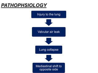
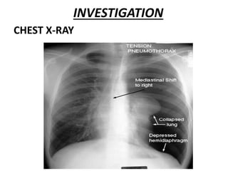
- 8. PATHOPHSIOLOGY Injury to the lung Valvular air leak Lung collapse Mediastinal shift to opposite side
- 9. Increased intrapleural pressure Decreased venous return Decreased ventilation Hypoxia and cardiac aarest
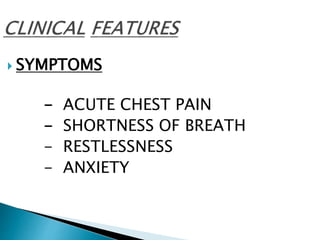
- 10. SYMPTOMS - ACUTE CHEST PAIN - SHORTNESS OF BREATH - RESTLESSNESS - ANXIETY
- 11. TACHYPNOEA CYANOSIS TACHYCARDIA JUGULAR VENOUS DISTENSION HYPOTENSION
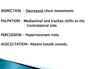
- 12. INSPECTION – Decreased chest movements PALPATION – Mediastinal and trachea shifts to the Contralateral side. PERCUSSION – Hyperresonant note. AUSCULTATION- Absent breath sounds.
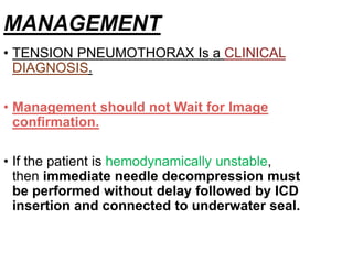
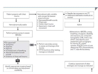
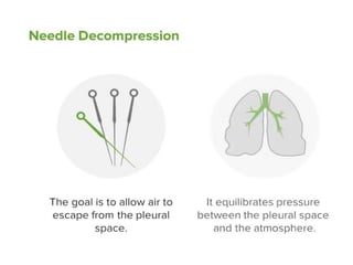
- 13. MANAGEMENT • TENSION PNEUMOTHORAX Is a CLINICAL DIAGNOSIS. • Management should not Wait for Image confirmation. • If the patient is hemodynamically unstable, then immediate needle decompression must be performed without delay followed by ICD insertion and connected to underwater seal.
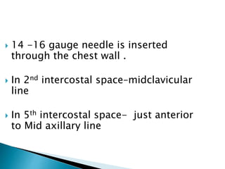
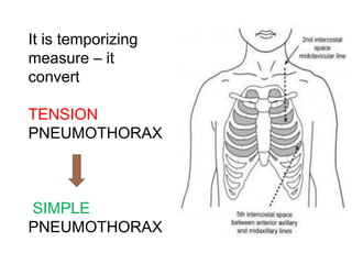
- 16. 14 -16 gauge needle is inserted through the chest wall . In 2nd intercostal space–midclavicular line In 5th intercostal space- just anterior to Mid axillary line
- 17. It is temporizing measure – it convert TENSION PNEUMOTHORAX SIMPLE PNEUMOTHORAX
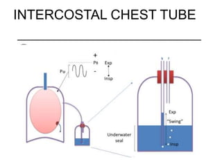
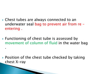
- 21. Chest tubes are always connected to an underwater seal bag to prevent air from re – entering . Functioning of chest tube is assessed by movement of column of fluid in the water bag . Position of the chest tube checked by taking chest X-ray
- 24. RESPIRATORY FAILURE RESPIRATORY ARREST CARDIAC ARREST SUBCUTANEOUS EMPHYSEMA PNEUMOPERICARDIUM PNEUMOPERITONEUM
- 25. PROCEDURE COMPLICATIONS Fistula formation Infections Bleeding Intercostal nerve injury
- 26. CARDIAC TAMPONADE HEMOTHORAX PULMONARY EMBOLISM MYOCARDIAL INFARCTION ACUTE AORTIC DISSECTION RIB FRACTURE COSTOCHONDRITIS DIAPHRAGMATIC INJURY
- 27. •BAILEY AND LOVE •SRB’S MANUAL OF SURGERY
- 28. THANK YOU

