This document discusses dyspepsia, defined as epigastric pain, burning, postprandial fullness, or early satiety. Dyspepsia can be caused by organic diseases like peptic ulcers, GERD, or malignancies. It can also be functional in nature. The evaluation of dyspepsia involves history, physical exam, and testing for H. pylori infection or structural abnormalities. Treatment depends on identified causes, but may include H. pylori eradication therapy, PPIs, or endoscopy.
Ulcerative colitis is a chronic inflammatory bowel disease that affects the colon. It is characterized by inflammation and ulcers in the lining of the rectum and colon. The causes are unknown but likely involve genetic and immune factors. Symptoms include abdominal pain, bloody diarrhea, and weight loss. Diagnosis involves blood tests, colonoscopy, and biopsy. Treatment focuses on reducing inflammation through medications like mesalamine, corticosteroids, immunosuppressants, or biologics. Surgery to remove the colon may be needed for severe cases or cancer prevention. Complications can include toxic megacolon, colon cancer, and extraintestinal manifestations.
This document summarizes acute and chronic gastritis. Acute gastritis is caused by factors like NSAID use, alcohol, smoking, infections, and stress. It involves neutrophil infiltration and can cause erosions and ulceration. Chronic gastritis is defined by long-term inflammation leading to atrophy and intestinal metaplasia. Helicobacter pylori infection is a major cause and results in urease production and cytotoxins that drive chronic inflammation. Autoimmune mechanisms can also lead to chronic gastritis, seen as lymphocytes and plasma cells in the lamina propria and intestinal metaplasia.
Systemic lupus erythematosus (SLE) is an autoimmune disease where the immune system attacks its own tissues, causing inflammation and damage. It is characterized by periods of disease flares and remission. Common symptoms include joint pain, rashes, and fatigue. SLE can affect many organs like the skin, lungs, heart, and kidneys. Diagnosis involves evaluating symptoms, lab tests like antinuclear antibodies, and sometimes biopsies. Treatment aims to reduce symptoms during flares and prevent organ damage using medications like corticosteroids, antimalarials, and immunosuppressants. SLE affects mostly women of childbearing age and has no known cure.
This document discusses gastritis, which is inflammation of the stomach lining. It defines acute and chronic gastritis and lists risk factors like smoking, drinking, and medications. Symptoms are discussed as well as diagnostic tests. Treatment focuses on antacids, bland diets, and treating any underlying causes. Chronic gastritis can lead to pernicious anemia or gastric cancer if not properly managed.
Irritable bowel syndrome (IBS) is a common functional gastrointestinal disorder characterized by abdominal pain and changes in bowel habits. The main symptoms are cramping, abdominal pain, bloating, gas, diarrhea and constipation. IBS has three subtypes based on predominant stool pattern: IBS with constipation, IBS with diarrhea, or IBS with mixed bowel habits. While the exact cause is uncertain, IBS involves abnormalities in gut motility and visceral sensitivity. Treatment involves lifestyle changes and medications to manage symptoms based on IBS subtype.
Chronic gastritis is defined as chronic inflammation of the stomach lining leading to atrophy and changes in cell type. The most common cause is infection by H. pylori bacteria, which causes damage through enzymes and toxins. This can lead to two types of gastritis - antral or pangastritis. Autoimmune gastritis is less common and is characterized by antibodies against stomach cells and vitamin B12 deficiency. H. pylori infection is generally treated with antibiotics and proton pump inhibitors to cure the infection and reduce cancer risk over the long term.
CHRONIC DYSPEPSIA
Seminar Prepared by :-
Ali Abdulazeem
Shilan Adnan Abdulrahman
Alaa Shamil
Guldan Hameed
Internal Medicine
College of Medicine - University of Kirkuk
This document provides an overview of irritable bowel syndrome (IBS), including its definition, prevalence, demographics, pathophysiology, clinical features, diagnosis, differential diagnosis, severity assessment, management, and prognosis. Some key points are:
- IBS is a functional bowel disorder characterized by abdominal pain associated with changes in bowel habits. It predominantly affects those aged 15-65 and is more common in women.
- The pathophysiology involves altered gut motility, visceral hypersensitivity, abnormal gas handling, low-grade inflammation, food sensitivities, abnormal gut microbiota, and central nervous system dysregulation.
- Diagnosis is based on symptoms meeting certain criteria and exclusion of organic diseases. Management focuses on
Cirrhosis is a late stage of scarring (fibrosis) of the liver caused by many forms of liver diseases and conditions, such as hepatitis and chronic alcoholism.
Clinical features and investigations of asthma is explained in very simple wording and style. Easy to remember and present due to interesting pictures. Helpful for medical students, patients with asthma and knowledge seekers.
Crohn's disease is an idiopathic inflammatory bowel disease characterized by transmural inflammation that can occur anywhere along the gastrointestinal tract from mouth to anus. It most commonly affects the terminal ileum and causes symptoms like diarrhea, abdominal pain, and weight loss. While the exact causes are unknown, it is believed to involve genetic susceptibility and an abnormal immune response triggered by environmental factors. Diagnosis involves examinations like colonoscopy, CT, MRI and blood tests. Treatment focuses on reducing inflammation using medications like antibiotics, aminosalicylates, corticosteroids, immunomodulators, and biologics. Complications can be intestinal, systemic, or postoperative.
Chronic glomerulonephritis is a kidney disorder caused by slow, cumulative damage and scarring of the glomeruli, or tiny blood filters in the kidneys, usually due to inflammation. This results in reduced kidney function over time and can lead to chronic kidney disease, end-stage renal disease, cardiovascular disease, renal failure, and death if left untreated. Treatment focuses on slowing disease progression, managing symptoms like high blood pressure and fluid retention, and renal replacement therapy with dialysis or kidney transplantation for kidney failure.
Esophagitis is inflammation of the esophagus that can have various causes like acid reflux, infections, medications, radiation, and more. Common symptoms include dysphagia, heartburn, and painful swallowing. Diagnosis involves endoscopy and biopsy. Treatment depends on the underlying cause but may include lifestyle changes, antacids, H2 blockers, proton pump inhibitors, and surgery in some cases. Complications can include strictures and Barrett's esophagus.
The document defines peptic ulcer disease as erosion of the GI mucosa from HCL acid and pepsin. Common sites are the lower esophagus, stomach, and duodenum. Risk factors include H. pylori infection, smoking, NSAIDs, and stress. Symptoms include epigastric pain, nausea, and tarry stools. Diagnosis involves endoscopy, blood tests, and breath tests for H. pylori. Treatment focuses on reducing acid with PPIs and antibiotics for H. pylori infection. Surgery may be needed for complications like bleeding or perforation. Long term management involves lifestyle changes and continued medication use.
Chronic gastritis is long-term inflammation of the stomach lining that can be caused by factors like H. pylori infection, medications, alcohol, or diet. It is classified based on etiology (cause), morphology (appearance), and location in the stomach. The OLGA staging system scores and stages gastritis severity based on the degree of atrophy in the antrum and corpus as seen histologically. Treatment involves eliminating the cause, such as treating an H. pylori infection, and managing symptoms.
This document discusses esophagitis, which is inflammation of the esophagus. It begins with an introduction defining esophagitis and describing its typical symptoms of painful swallowing and chest pain. It then covers the anatomy and histology of the esophagus, along with the various causes of esophagitis including acid reflux, infections, medications, and allergies. The document grades the severity of esophagitis and lists potential complications if left untreated, such as narrowing or abnormal tissue growth in the esophagus. It describes tests and diagnosis of esophagitis including barium X-rays, endoscopy, and biopsies. Treatment options involve medications to reduce acid or treat infections,
This document discusses peptic ulcers, including their causes, symptoms, diagnosis, and treatment. Peptic ulcers are abnormalities in the gastrointestinal tract caused by damage from stomach acid. The most common causes are infection with Helicobacter pylori bacteria and long-term use of nonsteroidal anti-inflammatory drugs. Common symptoms include abdominal pain, nausea, and vomiting of blood. Diagnosis involves tests to detect H. pylori infection and endoscopy to view the ulcers. Treatment focuses on eradicating H. pylori with antibiotics, reducing stomach acid with proton pump inhibitors or H2 blockers, and protecting the lining with sucralfate.
Gastro esophageal Reflux Disease (GERD) and its managementDr. Ankit Gaur
In this presentation I have tried to explain in brief about gastro esophageal Reflux Disease (GERD), its etiology, risk factors, diagnosis, and its management via pharmacotherapy.
This document provides an overview of cirrhosis of the liver. It begins by defining cirrhosis as the end stage of chronic liver disease, marked by diffuse involvement and disruption of the liver architecture with formation of nodules separated by fibrous bands. The causes of cirrhosis include alcoholic liver disease, viral hepatitis, genetic disorders. Clinically, cirrhosis presents with symptoms such as jaundice, ascites, peripheral edema and complications include hepatic coma, gastrointestinal bleeding and liver cancer. The progression of alcoholic liver disease from fatty liver to alcoholic hepatitis and finally cirrhosis is described along with the pathological changes at each stage.
This document provides information on evaluating and diagnosing acute and chronic abdominal pain. It discusses the history, physical exam, diagnostic studies, and management of various acute conditions like appendicitis, diverticulitis, cholecystitis, and perforated ulcer. It also covers chronic pain syndromes like irritable bowel syndrome and chronic pancreatitis. The goal is to distinguish between organic and functional causes of abdominal pain.
This document provides information on irritable bowel syndrome (IBS), including its definition, epidemiology, etiology, clinical features, diagnosis, investigations, treatment, and prognosis. IBS is a functional bowel disorder characterized by abdominal pain or discomfort and altered bowel movements in the absence of structural abnormalities. It has a prevalence of 1-20% worldwide and is more common in women. The cause is uncertain but may involve GI motor abnormalities, visceral hypersensitivity, brain-gut axis dysregulation, and abnormal psychology. Diagnosis is based on symptoms and ruling out other diseases. Treatment involves diet modification, pharmacotherapy including antispasmodics, antidepressants, and probiotics, as well as psychological therapies like CBT.
This document provides information on ascites including its definition, causes, diagnosis, and management. Ascites is defined as the accumulation of free fluid in the peritoneal cavity, most often caused by liver cirrhosis (75% of cases), malignancy, or heart failure. Diagnosis involves history, physical exam finding shifting dullness or fluid wave, and abdominal ultrasound or paracentesis. Initial ascites management consists of sodium restriction, diuretics, and large volume paracentesis for refractory ascites.
Irritable bowel syndrome (IBS) is a functional bowel disorder characterized by abdominal pain or discomfort associated with changes in bowel habits. IBS has no identifiable organic cause and is diagnosed based on symptom criteria. While IBS negatively impacts quality of life, it does not increase risk of serious disease or mortality. Potential contributing factors include abnormal gut motility, visceral hypersensitivity, disturbed pain processing, and psychiatric comorbidities like anxiety and depression. Differential diagnoses that require exclusion include inflammatory bowel disease, celiac disease, and colon cancer. All IBS patients should undergo basic blood tests and stool tests to rule out other conditions.
Chronic hepatitis is hepatic inflammation lasting at least 6 months caused by viruses, bacteria, toxins, or autoimmune conditions. It results in liver inflammation, damage, and fibrosis. Common symptoms include jaundice, abdominal pain, fatigue, and itching. Diagnosis involves blood tests showing liver enzyme elevations and liver biopsy demonstrating inflammation and fibrosis. Treatment focuses on addressing the underlying cause, diet, and medications like interferon for viral hepatitis. Chronic hepatitis can progress to cirrhosis if not properly managed.
This document provides information on dyspepsia, including its definition, causes, investigations, and management guidelines. It begins by defining dyspepsia and outlining its prevalence in the UK population. It then discusses the common and rare causes of dyspepsia and how to investigate patients. The document reviews guidelines from NICE on investigating and managing dyspepsia. It provides examples of case histories and questions to help apply the guidelines. Key points are emphasized, such as addressing lifestyle factors, empirically treating dyspepsia, and referring patients with red flag symptoms urgently for endoscopy.
Dyspepsia refers to any symptoms thought to originate from the upper gastrointestinal tract. There are several potential mechanisms that can cause dyspepsia, including gastroesophageal acid reflux, gastric motor dysfunction, and visceral afferent hypersensitivity. Gastroesophageal acid reflux can be caused by reduced lower esophageal sphincter tone, frequent transient lower esophageal sphincter relaxations, overeating, aerophagia, impaired esophageal body motility, reduced salivary secretion, and hiatal hernias. Gastric motor dysfunction may involve delayed gastric emptying or impaired gastric fundus relaxation after eating. Visceral afferent hypersensitivity is proposed to disturb gastric sensory function in functional
Helicobacter pylori infection and use of non-steroidal anti-inflammatory drugs (NSAIDs) are the two most common causes of peptic ulcer disease (PUD). H. pylori infection, found in 70-80% of PUD cases, causes chronic gastritis and impairs the stomach's defenses, allowing acid to damage the lining. NSAIDs also impair defenses and can cause ulcers even in the absence of H. pylori. Together these factors disrupt the stomach's balance between aggressive acid and pepsin secretions and protective mucosal defenses, leading to ulcer formation.
This document provides information on the causes, diagnosis, and management of peptic ulcer disease and gastric outlet obstruction. It discusses that peptic ulcers are caused by an imbalance between aggressive factors like acid secretion and defensive factors. Infection with H. pylori is a key cause. Diagnosis involves endoscopy with biopsy. Management focuses on treating H. pylori infection and using antisecretory drugs. Surgery is reserved for complications or refractory cases. Gastric outlet obstruction is usually due to peptic ulcer disease but can also be caused by malignancy, inflammation, or other conditions.
- Gastric and duodenal disorders like gastritis and peptic ulcers are common gastrointestinal problems that can be caused by factors like H. pylori infection, NSAID use, stress, and diet. Chronic gastritis and ulcers require long-term management like antibiotics, proton pump inhibitors, and lifestyle modifications.
- Peptic ulcers form in the stomach or duodenum due to an imbalance between gastric acid and the mucosal protective factors. Duodenal ulcers are more common than gastric ulcers and have distinguishing clinical features. Treatment aims to eliminate H. pylori and reduce acid with medications, surgery if needed.
- Morbid obesity is defined as being over 100 pounds
Ulcers range from small, painful sores in the mouth to bedsores and serious lesions of the stomach or interstine
Gastric ulcers :are peptic ulcers in the stomach.
Duodenal ulcers :are peptic ulcers in the duodenum
A peptic ulcer is a break in the stomach or duodenal lining that extends into deeper layers. Helicobacter pylori (H. pylori) infection and NSAID use are the most important risk factors. Common symptoms include recurrent epigastric pain relieved by food or antacids. Endoscopy is required for diagnosis and management. Eradication of H. pylori using PPIs and antibiotics is recommended to promote healing and prevent complications like bleeding. Surgery is only required for complications when medical management fails.
This presentation is about Peptic Ulcer Disease. I presented it in 2017 to my colleagues at Al Ain hospital. Information provided is up to date. I allow you to use it for educational purposes.
1. The document discusses chronic epigastric pain, its causes, symptoms, and methods of investigation and treatment. Common causes mentioned include gallstones, peptic ulcers, pancreatitis, and gastric carcinoma.
2. Diagnosis involves history, physical exam, and endoscopy to identify the specific cause. Treatment depends on the underlying condition but may include lifestyle changes, medications like PPIs, and eradication of H. pylori infection.
3. Surgery was previously used more often to treat peptic ulcers but has become less common with the availability of effective medical therapies. Surgical options described include various vagotomy procedures and gastrojejunostomy.
This document discusses peptic ulcer disease (PUD), including its causes, types, symptoms, diagnosis, and treatment. PUD is characterized by erosion of the GI mucosa from stomach acid and pepsin. It commonly affects the lower esophagus, stomach, and duodenum. The two main types are gastric and duodenal ulcers. Symptoms include abdominal pain, nausea, and vomiting. Diagnosis involves endoscopy and tests for H. pylori bacteria. Treatment focuses on reducing stomach acid with PPIs or H2 blockers, eradicating H. pylori, and protecting the mucosa. Complications can include bleeding, perforation, and obstruction if not properly treated.
Peptic ulcers are lesions that occur in areas of the gastrointestinal tract exposed to stomach acid. Risk factors include H. pylori infection and NSAID use. Clinical features include recurrent abdominal pain related to food. Diagnosis involves endoscopy with biopsy or breath/stool tests for H. pylori. Management involves eradicating H. pylori with triple therapy antibiotics and PPIs. Surgery is rarely needed and reserved for complications like perforation or bleeding.
peptic ulcer advance concepts of nursing.pptxajadoon84
The document discusses peptic ulcer disease including its definition, causes, symptoms, diagnostic testing, treatment, and nursing management. Peptic ulcers develop in the stomach, duodenum, or esophagus due to an imbalance between gastric acid and mucosal resistance. Common causes include H. pylori infection, NSAID use, and excess acid secretion. Symptoms include abdominal pain relieved by food or antacids as well as potential complications like bleeding. Treatment involves eradicating H. pylori, reducing acid production, and educating patients on lifestyle modifications.
This document discusses interventions for stomach disorders including gastritis, peptic ulcer disease, Zollinger-Ellison syndrome, and gastric cancer. It begins with an introduction to the anatomy and physiology of the stomach. Gastritis is then defined and the types, risk factors, pathogenesis, clinical features, diagnostic evaluation, and management are outlined. Peptic ulcer disease is similarly defined and the classifications, risk factors, etiological factors, pathogenesis, signs and symptoms, complications, diagnosis, and medical and non-medical management are described.
This document provides information on complications of peptic ulcer disease including perforation, hemorrhage, and gastric outlet obstruction. It discusses the pathophysiology, symptoms, diagnosis, and treatment options for these complications both conservatively and surgically. Surgical treatments covered include excision of ulcer with pyloroplasty, various types of vagotomy, and gastrectomy procedures.
Peptic ulcer disease is a break in the gastrointestinal mucosa caused by aggressive acid-peptic juices. Common sites are the stomach and duodenum. H. pylori infection and NSAID use are major causes. Clinical features include abdominal pain, nausea, and vomiting. Diagnosis involves endoscopy, biopsy, and H. pylori testing. Treatment includes lifestyle modifications, medications like PPIs, and H. pylori eradication therapy. Surgery is considered if complications develop or medical management fails. Proper counseling helps patients understand diet and medication.
Peptic Ulcer Disease is caused by breaks in the gastric or duodenal mucosa that are usually due to an imbalance between aggressive factors like acid and pepsin and protective ones like mucus and bicarbonate. H. pylori infection and NSAID use are major causes. Patients present with epigastric pain relieved by food or antacids. Diagnosis is usually made with endoscopy and tests for H. pylori. Treatment involves acid suppression, eradicating H. pylori with antibiotic combinations, and sometimes surgery for complications. Zollinger-Ellison Syndrome is a rare cause of ulcers due to a gastrin-secreting tumor.
Peptic ulcer disease is caused by an imbalance between gastric acid and mucosal defenses, allowing acid to damage the stomach or duodenum lining. Helicobacter pylori infection and NSAID use are leading causes. Symptoms include abdominal pain, nausea, and vomiting. Complications include bleeding, perforation, and cancer. Treatment involves eradicating H. pylori, stopping NSAIDs, and using PPIs to reduce acid and promote healing. Endoscopic therapies can help stop bleeding.
The document discusses diseases of the stomach, including stomach ulcers and dyspepsia. It provides details on the causes of stomach ulcers such as Helicobacter pylori bacteria and NSAIDs. Symptoms, complications, diagnosis and treatment are described for stomach ulcers. Dyspepsia is defined as indigestion and its common causes include diet, lifestyle factors, and underlying diseases. Treatment focuses on eliminating H. pylori infections, reducing stomach acid production, and managing symptoms.
This document discusses peptic ulcer disease, focusing on duodenal ulcers. It defines peptic ulcers and describes the pathogenesis, including protective and damaging factors. Helicobacter pylori infection plays a major role in ulcer development and the mechanisms by which it causes injury are explained. Diagnosis involves endoscopy, biopsy and testing for H. pylori. Treatment involves eradicating H. pylori with antibiotics and proton pump inhibitors. Complications of duodenal ulcers like bleeding and perforation are discussed. Bleeding ulcers are classified by the Forrest system and managed initially with endoscopic methods or surgery depending on severity. Perforated ulcers require surgical repair.
Peptic ulcer disease is defined as erosions in the gastric or duodenal mucosa that extend through the muscularis mucosae. Lifetime prevalence of peptic ulcer disease is 10% of Americans. Common causes include H. pylori infection, NSAID use, smoking, and alcohol consumption. Diagnosis involves history, physical exam, upper endoscopy with biopsy, and tests for H. pylori. Treatment focuses on eradicating H. pylori, reducing acid with PPIs, and lifestyle changes. Complications include bleeding, perforation, and gastric outlet obstruction.
Helicobacter pylori is a spiral-shaped bacteria that lives in the stomach. It was originally considered that bacteria could not survive in the acidic stomach. Marshall and Warren first cultured H. pylori from human stomachs and showed it was associated with gastric inflammation. H. pylori infection can cause dyspepsia, peptic ulcers, gastric cancer, and other diseases. Testing and treating H. pylori infections can help prevent future illness.
This document provides information on peptic ulcer disease, including its prevalence, risk factors, types, clinical manifestations, diagnosis, medical and surgical management, complications, nursing care, and follow up. Some key points:
- Peptic ulcers affect 4-10 per 1000 people in India and are more common in males aged 30-60. Risk factors include H. pylori infection, smoking, alcohol, NSAIDs.
- Types include acute, chronic, gastric, and duodenal ulcers. Chronic ulcers erode through the stomach/duodenal wall.
- Symptoms include abdominal pain relieved by food. Tests include endoscopy, biopsy to detect H. pylori.
- Treatment
Peptic ulcers are erosions in the stomach or duodenum caused by an imbalance between gastric acids and mucosal defenses. Risk factors include H. pylori infection, NSAIDs, smoking, and stress. H. pylori infection is the leading cause and eradication treatment involves PPIs and antibiotics. Complications of peptic ulcers include bleeding, perforation, and obstruction. Endoscopy is the best diagnostic tool and allows for treatment of bleeding ulcers. Surgery may be needed for complications or intractable disease.
Neck swelling - History taking, Causes, ClassificationTty Lim
This document provides guidance on evaluating neck swellings, including taking a history and performing an examination. It discusses assessing when the lump was first noticed, any associated symptoms, past medical history, and risk factors. A physical exam evaluates the lump's location, size, mobility, and texture. Potential causes of neck swellings include inflammatory/infectious processes, neoplasms, congenital/developmental abnormalities, and other rare entities. Further testing may be needed to arrive at a diagnosis and guide treatment.
Gout is a rheumatic disease caused by elevated levels of uric acid (hyperuricemia) which leads to the deposition of urate crystals in the joints. It predominantly affects males between 30-60 years of age and females after menopause. The prevalence is higher in developed countries and Oceanic populations. Hyperuricemia occurs due to increased production or decreased excretion of uric acid and causes an imbalance that results in crystal formation and inflammation in the joints.
Pleomorphic adenoma is the most common benign salivary gland tumor, arising most commonly in the parotid gland. It typically presents as a slow-growing, painless, firm, nodular mass in patients aged 30-40 years old. On histology, it demonstrates a biphasic appearance with both epithelial and myoepithelial cells within a variable stromal component. Diagnosis is made through imaging such as ultrasound, CT or MRI followed by surgical excision via enucleation or parotidectomy. Adenolymphoma, also known as Warthin's tumor, most commonly arises in the tail of the parotid gland in older males, appearing as an encapsulated cystic n
Wheeze is a high-pitched whistling sound caused by airflow moving through partially obstructed airways. It is produced when air passes through narrowed portions of the airways at high velocity, causing the airway walls to vibrate and alternately flatten and reopen. This vibration creates a continuous musical sound. Wheeze can be caused by conditions that narrow the airways such as asthma, bronchitis, pneumonia, and foreign body obstruction.
The document discusses laboratory testing of blood products. It covers:
1) Testing all blood donations for ABO group, RhD type, antibodies, and infections to ensure compatibility and safety.
2) Components of blood like red blood cells, platelets, plasma, and derivatives prepared from blood to treat specific conditions.
3) Procedures for blood grouping, antibody screening and identification, and compatibility testing to match safe blood to recipients.
This document describes forceps-assisted deliveries. It defines obstetric forceps as a double-bladed metal instrument used to extract the fetal head. It describes the parts of forceps including the blades, shanks, locks and handles. It discusses different types of forceps and their uses. It outlines the indications, prerequisites, technique and contraindications for a forceps-assisted delivery. Key steps include inserting the blades one at a time, applying traction in line with uterine contractions to deliver the baby. Training and experience of the operator are important to minimize risks.
HISTOLOGY OF ADRENAL GLAND & CORRELATION WITH FUNCTIONTty Lim
The adrenal gland has an outer cortex and inner medulla. The cortex is divided into three concentric zones - the zona glomerulosa secretes mineralocorticoids, the zona fasiculata secretes glucocorticoids, and the zona reticulari secretes androgens. The medulla contains chromaffin cells that secrete catecholamines such as epinephrine. Diseases can result from insufficient or excessive secretion from the cortex or medulla.
Effective communication among healthcare workersTty Lim
This document discusses effective communication among healthcare workers and ways to improve it. It contains the following key points:
- Poor communication is a major factor in medical errors, responsible for over 70% of sentinel events according to the Joint Commission. Medication errors and adverse drug effects are often caused by failures in team communication.
- Effective communication in healthcare is defined as a reciprocal, interactive process between sender and recipient to ensure information is received and understood. It is important for accurate diagnosis, detecting patient distress, and gaining patient satisfaction and compliance.
- Ways to improve individual communication abilities include developing assertiveness, active listening, and negotiation skills to challenge decisions respectfully and improve patient safety. Reducing distractions and adopting
This document discusses drug abuse, dependence, and addiction. It defines drug abuse as using drugs outside of social norms, while dependence occurs when abstaining from a drug causes withdrawal symptoms. Addiction means a drug dominates one's motivation. The document also discusses tolerance, psychological dependence, and physical dependence as the three basic processes of drug addiction. It provides statistics on drug use in Malaysia and lists common drug types like stimulants, depressants, hallucinogens, and opiates. Risk and protective factors for drug addiction are mentioned. References are included at the end.
This document discusses bilirubin metabolism and the causes of jaundice. It covers:
1) How bilirubin is normally produced from the breakdown of red blood cells and eliminated from the body through the liver and bile.
2) Causes of jaundice like hepatitis, bile obstruction, and hemolytic anemia which disrupt the normal balance of bilirubin production and clearance.
3) The pathology of cholestasis or impaired bile flow, including characteristic lab findings, morphological changes in the liver, and ductular proliferation in response to bile stasis.
The cerebral circulation receives 15% of cardiac output and autoregulates blood flow between arterial pressures of 60-140 mmHg. It is highly sensitive to changes in carbon dioxide, oxygen, hydrogen ion concentration, and sympathetic nervous activity to maintain neuronal homeostasis. The brain's blood supply is protected by collateral circulation through the Circle of Willis and intracranial pressure regulation according to the Monro-Kellie doctrine.
This document discusses systemic lupus erythematosus (SLE), a chronic autoimmune disease characterized by the formation of autoantibodies. SLE can affect many parts of the body including the skin, joints, heart, lungs, kidneys, and nervous system. Diagnosis involves detecting antinuclear antibodies (ANAs) and other autoantibodies in the blood through tests like the fluorescent antinuclear antibody (FANA) test. FANA testing can identify different antibody patterns that may indicate SLE or other autoimmune conditions. Laboratory tests are also used to monitor disease activity and screen for organ involvement.
Upper respiratory tract infections are very common, usually manifesting as the common cold with symptoms of nasal congestion, watery discharge, sneezing, and sore throat. The most common pathogens are rhinoviruses, coronaviruses, and influenza viruses. Acute infections can also involve the pharynx, larynx, and epiglottis, sometimes leading to complications like bacterial infections. Nasopharyngeal carcinoma is a rare cancer linked to Epstein-Barr virus that invades locally and spreads to lymph nodes and distant sites. Laryngeal tumors include vocal cord nodules, papillomas, and squamous cell carcinomas, with the latter being the most significant due to its link
Skeletal Muscle Fatigue and Cellular MechanismsTty Lim
Muscle fatigue is a decline in muscle performance associated with activity that is temporary and reversible. It occurs due to fuel depletion, buildup of metabolic byproducts, and neuromuscular events. Specifically, fatigue results from depletion of ATP and glycogen stores, accumulation of hydrogen ions and inorganic phosphate, and impaired sodium and potassium gradients. This leads to reduced force generation, calcium sensitivity, and calcium release. Fatigue is also caused by acidosis as hydrogen ions compete with calcium ions. Recovery from fatigue involves rest, active recovery like massage, replenishing fuel stores through diet, and rehydration.
Plasma derived chemical mediators of inflammation - ttylimTty Lim
The document describes three plasma protein-derived mediator systems - the complement, kinin, and coagulation systems. These systems consist of plasma proteins that play important roles in host defense and inflammation. The complement system includes proteins C1-C9 that are activated by proteolysis in a cascade. The kinin system involves Hageman factor, high molecular weight kininogen, bradykinin, and kallikrein. The coagulation system includes thrombin, fibrinogen, and fibrinopeptides. Mediators from these systems such as C3a, C5a, bradykinin, and thrombin induce inflammation through vascular effects, leukocyte activation, and phagocytosis.
Chair, Joshua Sabari, MD, discusses NSCLC in this CME activity titled “Modern Practice Principles in Lung Cancer—First Find the Targets, Then Treat With Precision: A Concise Guide for Biomarker Testing and EGFR-Targeted Therapy in NSCLC.” For the full presentation, downloadable Practice Aid, and complete CME information, and to apply for credit, please visit us at https://bit.ly/3VomnBV. CME credit will be available until February 26, 2026.
Chair, Grzegorz (Greg) S. Nowakowski, MD, FASCO, discusses diffuse large B-cell lymphoma in this CME activity titled “Addressing Unmet Needs for Better Outcomes in DLBCL: Leveraging Prognostic Assessment and Off-the-Shelf Immunotherapy Strategies.” For the full presentation, downloadable Practice Aid, and complete CME information, and to apply for credit, please visit us at https://bit.ly/49JdxV4. CME credit will be available until February 27, 2026.
Creatine’s Untold Story and How 30-Year-Old Lessons Can Shape the FutureSteve Jennings
Creatine burst into the public consciousness in 1992 when an investigative reporter inside the Olympic Village in Barcelona caught wind of British athletes using a product called Ergomax C150. This led to an explosion of interest in – and questions about – the ingredient after high-profile British athletes won multiple gold medals.
I developed Ergomax C150, working closely with the late and great Dr. Roger Harris (1944 — 2024), and Prof. Erik Hultman (1925 — 2011), the pioneering scientists behind the landmark studies of creatine and athletic performance in the early 1990s.
Thirty years on, these are the slides I used at the Sports & Active Nutrition Summit 2025 to share the story, the lessons from that time, and how and why creatine will play a pivotal role in tomorrow’s high-growth active nutrition and healthspan categories.
TunesKit Spotify Converter Crack With Registration Code 2025 Freedfsdsfs386
TunesKit Spotify Converter is a software tool that allows users to convert and download Spotify music to various formats, such as MP3, AAC, FLAC, or WAV. It is particularly useful for Spotify users who want to keep their favorite tracks offline and have them in a more accessible format, especially if they wish to listen to them on devices that do not support the Spotify app.
https://shorturl.at/LDQ9c
Copy Above link & paste in New Tab
Dr. Jaymee Shell’s Perspective on COVID-19Jaymee Shell
Dr. Jaymee Shell views the COVID-19 pandemic as both a crisis that exposed weaknesses and an opportunity to build stronger systems. She emphasizes that the pandemic revealed critical healthcare inequities while demonstrating the power of collaboration and adaptability.
Shell highlights that organizations with gender-diverse executive teams are 25% more likely to experience above-average profitability, positioning diversity as a business necessity rather than just a moral imperative. She notes that the pandemic disproportionately affected women of color, with one in three women considering leaving or downshifting their careers.
To combat inequality, Shell recommends implementing flexible work policies, establishing clear metrics for diversity in leadership, creating structured virtual collaboration spaces, and developing comprehensive wellness programs. For healthcare providers specifically, she advocates for multilingual communication systems, mobile health units, telehealth services with alternatives for those lacking internet access, and cultural competency training.
Shell emphasizes the importance of mental health support through culturally appropriate resources, employee assistance programs, and regular check-ins. She calls for diverse leadership teams that reflect the communities they serve and community-centered care models that address social determinants of health.
In her words: "The COVID-19 pandemic didn't create healthcare inequalities – it illuminated them." She urges building systems that reach every community and provide dignified care to all.
BIOMECHANICS OF THE MOVEMENT OF THE SHOULDER COMPLEX.pptxdrnidhimnd
The shoulder complex acts as in coordinated fashion to provide the smoothest and greatest range of motion possible of the upper limb.
Combined motion of GH and ST joint of shoulder complex helps in:
Distribution of motion between other two joints.
Maintenance of glenoid fossa in optimal position.
Maintenance of good length tension
Although some amount of glenohumeral motion may occur while the other shoulder articulations remain stabilized, movement of the humerus more commonly involves some movement at all three shoulder joints.
Digestive Powerhouses: Liver, Gallbladder, and Pancreas for Nursing StudentsViresh Mahajani
This educational PowerPoint presentation is designed to equip GNM students with a solid understanding of the liver, pancreas, and gallbladder. It explores the anatomical structures, physiological processes, and clinical significance of these vital organs. Key topics include:
Liver functions: detoxification, metabolism, and bile synthesis.
Gallbladder: bile storage and release.
Pancreas: exocrine and endocrine functions, including digestive enzyme and hormone production. This presentation is ideal for GNM students seeking a clear and concise review of these important digestive system components."
This presentation provides a detailed exploration of the morphological and microscopic features of pneumonia, covering its histopathology, classification, and clinical significance. Designed for medical students, pathologists, and healthcare professionals, this lecture differentiates bacterial vs. viral pneumonia, explains lobar, bronchopneumonia, and interstitial pneumonia, and discusses diagnostic imaging patterns.
💡 Key Topics Covered:
✅ Normal lung histology vs. pneumonia-affected lung
✅ Morphological changes in lobar, bronchopneumonia, and interstitial pneumonia
✅ Microscopic features: Fibroblastic plugs, alveolar septal thickening, inflammatory cell infiltration
✅ Stages of lobar pneumonia: Congestion, Red hepatization, Gray hepatization, Resolution
✅ Common causative pathogens (Streptococcus pneumoniae, Klebsiella pneumoniae, Mycoplasma, etc.)
✅ Clinical case study with diagnostic approach and differentials
🔬 Who Should Watch?
This is an essential resource for medical students, pathology trainees, and respiratory health professionals looking to enhance their understanding of pneumonia’s morphological aspects.
2.
defined as having one or more symptoms of
epigastric pain, burning, postprandial fullness, or
early satiety.
Bloating
Nausea
Loss of appetite
Dyspepsia
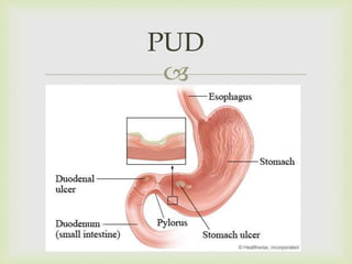
4.
Type I
typically located near the angularis incisura on the lesser
curvature, close to the border between the antrum and the
body of the stomach. Patients with type I gastric ulcers
usually have normal or decreased gastric acid secretion.
Type II
a combination of stomach and duodenal ulcers and are
associated with normal or increased gastric acid secretion.
Type III
prepyloric and are associated with normal or increased
gastric acid secretion.
Type IV
occur near the gastroesophageal junction, and gastric acid
secretion is normal or below normal.
Types of Gastric Ulcer
6. H. pylori
70% of gastric ulcer patients are infected with H. pylori.
Majority of colonised people remain healthy and
asymptomatic.
uses adhesin molecules (BabA) to bind to Lewis b antigen on
epeithelial cells.
induces an intense inflammatory and immune response
IL-1, IL-6, tumor necrosis factor, IL-8
Production of ammonia by the enzyme urease
Toxic to epithelial cell
Increase gastrin release from G cells
Negative feedback loop for gastrin release is halted
stimulates increased acid production by parietal cells.
Pathophysiology
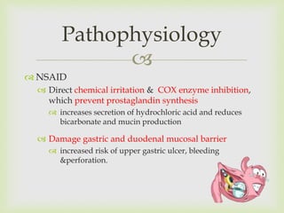
7.
NSAID
Direct chemical irritation & COX enzyme inhibition,
which prevent prostaglandin synthesis
increases secretion of hydrochloric acid and reduces
bicarbonate and mucin production
Damage gastric and duodenal mucosal barrier
increased risk of upper gastric ulcer, bleeding
&perforation.
Pathophysiology
8.
Smoking
Increased risk of gastric ulcer and duodenal ulcer to a
lesser extent.
more likely to causing complication and less likely to
heal if the patient continues to smoke.
Pathophysiology
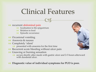
9.
recurrent abdominal pain
localisation to the epigastrium
Relation to food
Episodic occurrance
Occasional vomiting
Anorexia & nausea
Completely ‘silent’
presented with anaemia for the first time
Recurrent acute bleeding without ulcer pain
Gnawing or burning sensation
occurs shortly after meals with gastric ulcer and 2-3 hours afterward
with duodenal ulcer.
Diagnostic value of individual symptoms for PUD is poor.
Clinical Features
10.
Upper GI endoscopy
Rapid urease tests
Fecal antigen testing
detecting the presence of H pylori antigens in stools
Urea breath test
testing for the enzymatic activity of bacterial urease.
Antibodies (IgG)
X- ray
detect free abdominal air when perforation is suspected.
upper GI contrast study
extravasation of contrast indicates gastric perforation
Investigation
11.
H. pylori eradication
proton pump inhibitor (PPI)–based triple therapy.
PPI, amoxicillin, and clarithromycin for 7-14 days.
Amoxicillin should be replaced with metronidazole in penicillin-
allergic patients only high rate of metronidazole resistance
NSAID
American College of Gastroenterology (ACG ) guideline: test
for H pylori done in patients who started long-term NSAID
therapy
NSAIDs should be immediately discontinued in patients with
positive H pylori test results if clinically feasible
Patient with known history of ulcer and in whom NSAID use is
unavoidable, the lowest possible dose and duration of the
NSAID and co-therapy with a PPI or misoprostol are
recommended.
Treatment
12.
Surgical
Rarely required
Choice for a chronic non-healing gastric ulcer
partial gastrectomy to exclude an underlying cancer.
Treatment
13.
Duodenal ulcer and gastric ulcer both belong to the
family of peptic ulcer disease.
H. pylori infection is the major cause of duodenal
ulcer followed by NSAID
They share almost the same clinical features.
Duodenal Ulcer
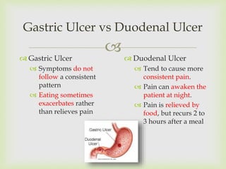
14.
Gastric Ulcer
Symptoms do not
follow a consistent
pattern
Eating sometimes
exacerbates rather
than relieves pain
Gastric Ulcer vs Duodenal Ulcer
Duodenal Ulcer
Tend to cause more
consistent pain.
Pain can awaken the
patient at night.
Pain is relieved by
food, but recurs 2 to
3 hours after a meal
16.
Harmon RC, Peura DA. Evaluation and Management of
Dyspepsia [Internet]. Medscape. [cited 2015 May 24]. Available
from: http://www.medscape.com/viewarticle/721062_1
Robbins basic Pathology. 9th Ed.
BS Anand. Peptic Ulcer Disease Treatment & Management
[Internet]. [cited 2015 May 24]. Available from:
http://emedicine.medscape.com/article/181753-
treatment#aw2aab6b6b1aa
Davidson’s Principle & Practice of Medicine. 22nd Ed.
References
Editor's Notes
#7: Although H. pylori does not invade the tissues, it induces an intense inflammatory and immune response. There is increased production of proinflammatory cytokines such as interleukin (IL)-1, IL-6, tumor necrosis factor, and, most notably, IL-8. IL-8 is produced by the mucosal epithelial cells, and it recruits and activates neutrophils.Several bacterial gene products are involved in causing epithelial cell injury and induction of inflammation. Epithelial injury is mostly caused by a vacuolating toxin called VacA, which is regulated by the cytotoxin-associated gene A (CagA). This gene is a component of the Cag pathogenicity island, a cluster of 29 genes, some of which encode pro-inflammatory proteins. In addition, H. pylori secretes a urease that breaks down urea to form toxic compounds such as ammonium chloride and monochloramine. The organisms also elaborate phospholipases that damage surface epithelial cells. Bacterial proteases and phospholipases break down the glycoprotein-lipid complexes in the gastric mucus, thus weakening the first line of mucosal defense.H. pylori enhances gastric acid secretion and impairs duodenal bicarbonate production, thus reducing luminal pH in the duodenum. This altered milieu seems to favor gastric metaplasia (the presence of gastric epithelium) in the first part of the duodenum. Such metaplastic foci provide areas for H. pylori colonization.Several H. pylori proteins are immunogenic, and they evoke a robust immune response in the mucosa. Both activated T cells and B cells can be seen in chronic gastritis caused by H. pylori. The B lymphocytes aggregate to form follicles. The role of T and B cells in causing epithelial injury is not established, but T-cell-driven activation of B cells may be involved in the pathogenesis of gastric lymphomas
#8: Suppression of mucosal prostaglandin synthesis, which increases secretion of hydrochloric acid and reduces bicarbonate and mucin production