The Human Eye
Human Eye And Colourful World of Class 10
The human eye is one of the most valuable and sensitive sense organs, which enable us to see the colourful world around us. It is just like a photographic camera. The lens system of the eye forms an image of the object on a light sensitive screen.
STRUCTURE AND WORKING OF HUMAN EYE:
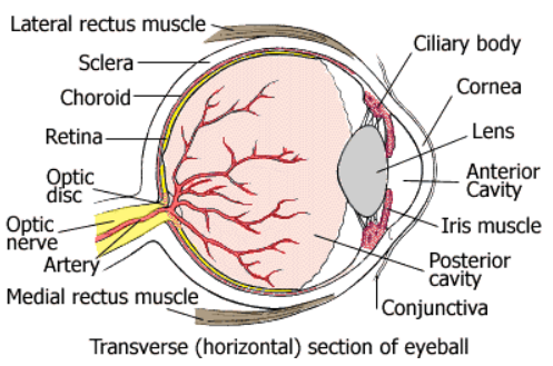
The human eye has the following parts :
- Cornea : It is the transparent spherical membrane covering the front of the eye.
- Iris : It is the coloured diaphragm between the cornea and lens.
- Pupil : It is the small hole in the iris.
- Eye lens : it is a transparent lens made of jelly like material.
- Ciliary muscles : These muscles hold the lens in positions.
- Retina : It is the back surface of the eye.
- Blind spot : It is the point at which the optic nerve leaves the eye. An image formed at this point is not sent to the brain.
- Aqueous humor : It is clear liquid region between the cornea and the lens.
- Vitreous humor : The space between eye lens and retina is filled with another liquid called vitreous humor.In the eye, the image is formed on the retina by successive refractions at the cornera, the aqueous humor, the lens and the vitreous humor. Electrical signals then travel along the optic nerve to the brain to be interpreted. In good light, the yellow spot is most sensitive to detail and the image is automatically formed there.
WORKING OF THE HUMAN EYE
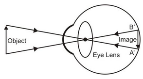
Light rays coming from the object to be seen can enter the eye through pupil and fall on the eye lens. The eye lens being convex, forms a real and inverted image on the retina. The light sensitive cell of the retina gets activated upon illumination and generate electric signals. These signals are sent to the brain via the optic nerve. The brain interprets these signals and finally gives rise to the sensation of vision.
THE FUNCTION OF IRIS AND PUPIL
The iris adjusts the size of the pupil according to the intensity of light received by the eye. The pupil regulates and controls the amount of light entering the eye. When the intensity of outside light is high, the pupil contracts so that less light enters the eye. When the intensity of the outside light is low, the pupil expands so that more light enters the eye and we can see properly.
Power of Accommodation:
The image of the objects at different distances from the eye are brought to focus on the retina by changing the focal length of the eye-lens, which is composed of fibrous jelly-like material, can be modified to some extent by the ciliary muscles.
Near Point and Far Point:
The nearest point at which a small object can be seen distinctly by the eye is called the near point. For a normal eye, it is about 25 cm and is denoted by the symbol D.
With advancing age, the power of accommodation of the eye decreases at the eye lens gradually loses its flexibility. For most of the old persons aged nearly 60 years, the near point is about 200 cm and corrective glasses are needed to see the nearby objects clearly.
The farthest point upto which our eye can seen objects clearly, without any strain on the eye is called the far point. For a person with normal vision, the far point is at infinity.
Least Distance of Distinct Vision:
The minimum distance of an object from the eye at which it can be seen most clearly and distinctly without any strain on the eye, is called the least distance of distance of distinct vision. For a person with normal vision, it is about 25 cm and is represented by the symbol D, i.e.
Least distance of distinct vision = D = 25 cm
Persistence of Vision:
The image formed on the retina of the eye does not fade away. Instantaneously, when the object is removed from the sight. The impression (or sensation) of the object remains on the retina for about (1/16)th of a second, even after the object is removed from the sight. This continuance of the sensation of eye is called the persistence of vision.
Let a sequence of still pictures is taken by a move camera. If the sequence of these still pictures is projected on a screen at a rate of 24 images or more per second then the successive impression of the images on the screen appear to blend or merge smoothly into one another. This is because an image (or a scene) on the screen appears just before the impression of previous image on the retina is lost. Hence, the sequence of images blend into one another giving the impression of a moving picture. This principle is used in motion picture projection or in cinematography
Colour - Blindness:
The retina of our eye has large number of light sensitive cells having shapes of rods and cones. The rod-shaped cells responds to the intensity of light with different of brightness and darkness were as the cone shaped cells respond to colour. In dim light rods are sensitive, but cones are sensitive only bright. The cones are sensitive to red, green and blue colour of light to different extents.
Due to genetic disorder, some persons do not possess some cone-shaped cells that responds to certain specific colours only. Such persons cannot distinguish between certain colour but can seen well otherwise. Such persons are said to have colour-blindness. Driving licenses are generally not issued to persons having colour-blindness.
Colour Perception of Animals:
Different animals have different colour perception due to which different structure of rod shaped cells and core shaped cells. For example, bees have some cone-shaped cells that are sensitive to ultraviolet. Therefore bees can seen objects in ultraviolet light and can perceive colours which we cannot do.
Human beings cannot seen in ultraviolet light as their retina do not have cone-shaped cells that are sensitive to ultraviolet light.
The retina of chicks have mostly cone shaped cells and only a few rod shaped cells. As rod shaped cells are sensitive to bright light only. Therefore, chicks wake up with sunrise and sleep in their resting place by the sunset.
Cataract:
Sometimes due to the formation of a membrane over the crystalline lens of some people in the old age, the eye lens becomes hazy or even opaque. This is called cataract. It results in decrease or loss in vision of the eye. Cataract can be corrected by surgery leading to normal vision.


